. 2010 Feb 17;30(7):2418-27.
doi: 10.1523/JNEUROSCI.5533-09.2010.

Hormonal modulation of sensorimotor integration

Affiliations

Hormonal modulation of sensorimotor integration

Nicholas D DeLong et al. J Neurosci. .

Abstract

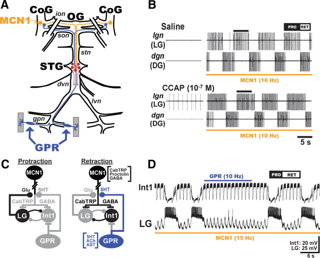
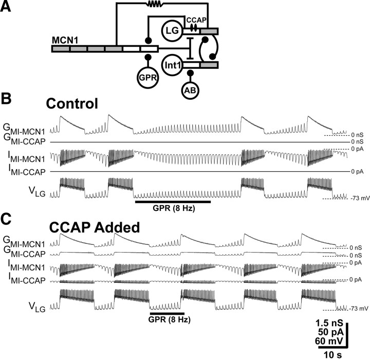
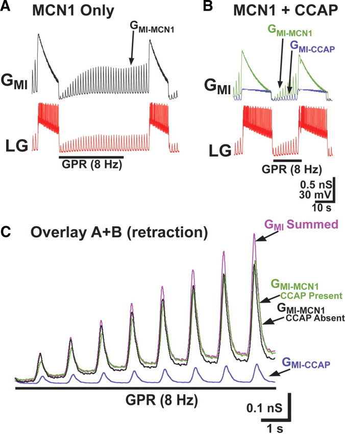
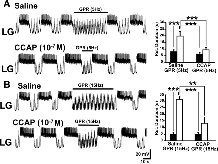
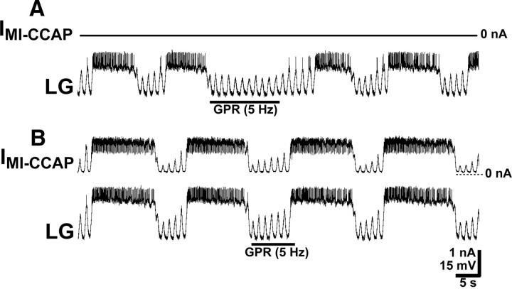
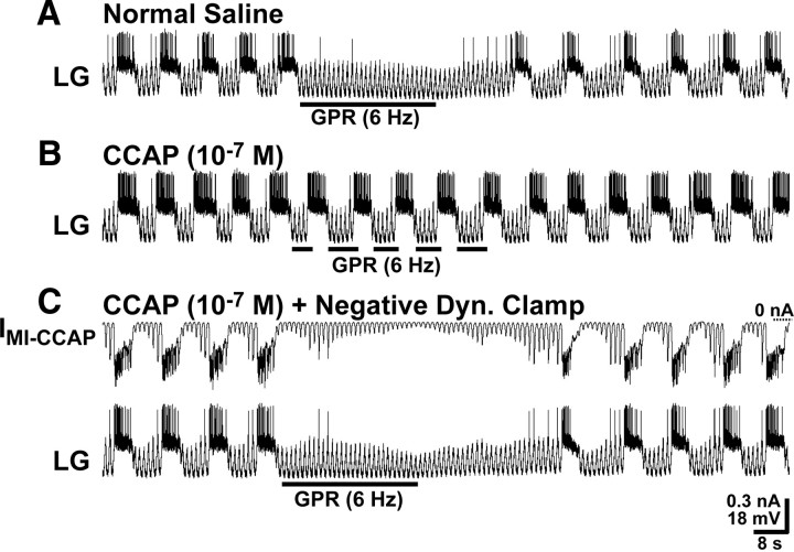
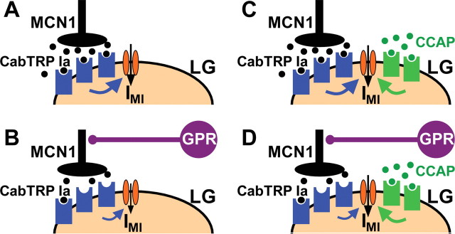
Neuronal circuits commonly receive simultaneous inputs from descending, ascending, and hormonal systems. Thus far, however, most such inputs have been studied individually to determine their influence on a given circuit. Here, we examine the integrated action of the hormone crustacean cardioactive peptide (CCAP) and the gastropyloric receptor (GPR) proprioceptor neuron on the biphasic gastric mill (chewing) rhythm driven by the projection neuron modulatory commissural neuron 1 (MCN1) in the isolated crab stomatogastric ganglion. In control saline, GPR stimulation selectively prolongs the gastric mill retractor phase, via presynaptic inhibition of MCN1. In the absence of GPR stimulation, CCAP does not alter retraction duration and modestly prolongs protraction. Here, we show, using computational modeling and dynamic-clamp manipulations, that the presence of CCAP weakens or eliminates the GPR effect on the gastric mill rhythm. This CCAP action results from its ability to activate the same modulator-activated conductance (G(MI)) as MCN1 in the gastric mill circuit neuron lateral gastric (LG). Because GPR prolongs retraction by weakening MCN1 activation of G(MI) in LG, the parallel G(MI) activation by CCAP reduces the impact of GPR regulation of this conductance. The CCAP-activated G(MI) thus counteracts the GPR-mediated decrease in the MCN1-activated G(MI) in LG and reduces the GPR ability to regulate the gastric mill rhythm. Consequently, although CCAP neither changes retraction duration nor alters GPR inhibition of MCN1, its activation of a modulator-activated conductance in a pivotal downstream circuit neuron enables CCAP to weaken or eliminate sensory regulation of motor circuit output.


Figures

Figure 1.
Figure 1.
Schematics of the isolated STNS and the MCN1-elicited gastric mill circuit, as well as its regulation by CCAP and GPR. A, In each CoG, there is a single copy of the projection neuron MCN1, which projects an axon through the ion and stn nerves to the STG. Each GPR projects an axon through the lvn and dvn nerves to arborize in the STG and continues through the stn and son nerves to innervate each CoG. The paired diagonal bars through the sons and ions represent the transection of these nerves at the start of each experiment. The gray rectangles represent protractor muscles in which the GPR dendrites arborize. These muscles were removed for the experiments in this paper. Abbreviations: Ganglia: CoG, commissural ganglion; OG, oesophageal ganglion. Nerves: dvn, dorsal ventricular nerve; lvn, lateral ventricular nerve; stn, stomatogastric nerve. B, As shown by Kirby and Nusbaum (2007), bath-applied CCAP selectively prolongs the protractor phase of the MCN1-elicited gastric mill rhythm. Note that CCAP did not activate the gastric mill rhythm before MCN1 stimulation. Protraction (PRO) phase is represented by the LG protractor neuron activity. Retraction (RET) phase is represented by the dorsal gastric (DG) retractor neuron activity. The bar on top of second LG burst in each panel represents the LG burst duration in saline, to show that the LG burst is prolonged by CCAP. This panel was reproduced from the study by DeLong et al. (2009a). C, Gastric mill CPG circuit schematics during each phase of the gastric mill rhythm. The paired diagonal bars through MCN1 axon represent additional distance between CoG and STG. All synapses shown are located in the STG. The gray somata and synapses represent neurons/synapses that are inactive during the indicated phase of the gastric mill rhythm. Synapses drawn on somata or axons actually occur on small branches in the STG neuropil. Transmitters in brackets next to MCN1 and GPR somata are their identified cotransmitters. Note that MCN1 uses only CabTRP Ia to excite LG and only GABA to excite Int1, whereas GPR uses only 5-HT to inhibit MCN1STG (Wood et al., 2000; Stein et al., 2007; DeLong et al., 2009b). LG and Int1 are both glutamatergic (Marder, 1987; Saideman et al., 2007a). Transmitter abbreviations: 5-HT, 5-hydroxytryptamine (serotonin); Glu, glutamate. Symbols: Filled circles, Synaptic inhibition; T-bars, synaptic excitation. D, GPR stimulation selectively prolongs the gastric mill retractor phase. Note that the duration of the retractor phase during GPR stimulation is longer than the same phase in the cycles immediately before and after GPR stimulation (Beenhakker et al., 2005). The fast rhythmic action potential bursts in Int1 and the associated fast rhythmic subthreshold LG oscillations represent the influence of the pyloric circuit on the gastric mill CPG (Bartos et al., 1999). The rhythmic subthreshold depolarizations in LG result from the rhythmic removal of Int1-mediated inhibition and the consequent unmasking of MCN1-mediated excitation. A, C, and D are reproduced from the study by DeLong et al. (2009b).
Figure 2.
Figure 2.
GPR regulation of the gastric mill retractor phase is weakened by the presence of CCAP in a computational model. A, Circuit schematic representing the CCAP modulation of the GPR influence on the MCN1-activated gastric mill CPG in a compartmental model. The model is modified from the original model in the study by Nadim et al. (1998) by the addition of the indicated CCAP and GPR actions. Specifically, as in previously published models, the CCAP-activated conductance was added to the LG neurite compartment (DeLong et al., 2009a), and an inhibitory GPR synapse was added onto the passive terminal compartment of MCN1 (Beenhakker et al., 2005; DeLong et al., 2009b). The gray compartments have active properties mediated by voltage-dependent, Hodgkin–Huxley-like conductances to facilitate action potential generation, whereas the white compartments are passive. Symbols: Filled circles, Synaptic inhibition; T-bars, synaptic excitation; resistor, electrical coupling. B, GPR selectively prolongs the retractor phase of the MCN1-elicited gastric mill rhythm in the absence of CCAP. Under these conditions, the MCN1-elicited inward current (IMI-MCN1) and associated conductance (GMI-MCN1) in LG grew in amplitude during each retractor (LG-silent) phase because of continual MCN1 release of CabTRP Ia, and decayed during each protractor (LG-active) phase because of the LG presynaptic inhibition of MCN1STG (Figs. 1C, 2A). As in the biological preparation, GPR stimulation during the retractor phase selectively prolonged that phase (Beenhakker et al., 2005; DeLong et al., 2009b). The fast transient events in IMI-MCN1 resulted from the rapid changes in driving force as the LG membrane potential repeatedly approached the IMI reversal potential during the LG action potentials (DeLong et al., 2009a). C, Adding the CCAP-activated conductance (GMI-CCAP) to LG reduced the ability of GPR to prolong retraction. In the presence of GMI-CCAP, the GPR stimulation was less effective in prolonging the retractor phase relative to the control condition (B). Note also that, as reported previously (Kirby and Nusbaum, 2007; DeLong et al., 2009b), GMI-MCN1 and GMI-CCAP exhibited different trajectories during the LG burst, and it is the sustained GMI-CCAP amplitude during protraction (LG burst) that prolonged this phase relative to the control condition (B). As above, the fast transient events in IMI-MCN1 and IMI-CCAP resulted from the rapid changes in driving force during the LG action potentials.
Figure 3.
Figure 3.
The altered GMI dynamics in the LG neuron when CCAP is present reduces the ability of GPR to prolong the gastric mill retractor phase in a computational model. A, During the control gastric mill rhythm, GMI in LG was entirely attributable to input from MCN1 (GMI-MCN1). Under these conditions, GPR stimulation prolonged the retractor phase (Fig. 2B) by reducing the rate of buildup of GMI-MCN1 caused by the GPR presynaptic inhibition of MCN1 (Beenhakker et al., 2005; DeLong et al., 2009b). B, When CCAP was present, GMI consisted of the summed components contributed by MCN1 (GMI-MCN1) and CCAP (GMI-CCAP). Under this condition, GPR stimulation was less effective in prolonging retraction. C, The presence of CCAP during MCN1 stimulation produced a summed maximal GMI in LG during the retractor phase that grew at a faster rate than when GMI was entirely contributed by MCN1. The GMI traces from A and B are overlaid, for durations that span the first ∼10 s of the retractor phase during GPR stimulation. With CCAP both present and absent, the amplitude of GMI-MCN1 grew steadily during retraction. However, the presence of CCAP produced a summed maximal GMI that grew at a faster rate, because it was additionally composed of the GPR-independent GMI-CCAP. Hence, the summed maximal GMI in the presence of CCAP enabled LG to attain burst threshold sooner.
Figure 4.
Figure 4.
CCAP superfusion reduces the effectiveness of GPR stimulation during the biological MCN1-elicited gastric mill rhythm. A, Left, GPR stimulation selectively prolonged retraction (LG silent) under control (saline) conditions during the MCN1–gastric mill rhythm. In contrast, the same level of GPR stimulation barely prolonged retraction during CCAP superfusion. Note that, as usual, CCAP prolonged LG burst duration (Kirby and Nusbaum, 2007). Most hyperpolarized Vm: saline, −62 mV; CCAP, −65 mV. Right, Cumulative data showing that GPR consistently prolonged the gastric mill retractor phase during saline superfusion. In contrast, GPR stimulation did not alter retraction duration in the presence of CCAP (10−7 m; RM-ANOVA, SNK post hoc test, p = 0.32; n = 5). The retraction duration was also prolonged by the GPR stimulation in saline compared with the same stimulation in the presence of CCAP, whereas there was no difference in the duration of this phase during the two control conditions (RM-ANOVA, SNK post hoc test, p = 0.99; n = 5). ***RM-ANOVA, SNK post hoc text, p < 0.001; n = 5. The black bars represent gastric mill cycles without GPR stimulation, and the white bars represent cycles with GPR stimulation. Error bars indicate SEM. B, Left, Increasing the GPR stimulation frequency prolonged the gastric mill retractor phase in saline but still failed to maintain its effectiveness during CCAP superfusion. Most hyperpolarized Vm: Both panels, −58 mV. Right, Cumulative data showing that increasing the GPR stimulation frequency prolongs the retractor phase during saline superfusion. However, this faster stimulation frequency was not sufficient to overcome the influence of CCAP (10−7 m) (RM-ANOVA, SNK post hoc test, p = 0.13; n = 3). Also, as in A, the retraction duration was distinct during the GPR stimulations in saline and CCAP, but not during the two control conditions (RM-ANOVA, SNK post hoc test, p = 0.90; n = 3). ***RM-ANOVA, SNK post hoc test, p < 0.001; **RM-ANOVA, SNK post hoc test, p < 0.005. The bars are as above.
Figure 5.
Figure 5.
Dynamic-clamp injection of the CCAP-activated current (IMI-CCAP) into LG is sufficient to mimic the ability of bath-applied CCAP to weaken the GPR action on the MCN1-elicited gastric mill rhythm. A, In the absence of the dynamic-clamp injection (IMI-CCAP, 0 nA), GPR stimulation during the gastric mill retractor phase selectively prolonged that phase. Most hyperpolarized Vm, −59 mV. B, Dynamic clamp depolarizing current injection of IMI-CCAP into LG mimicked the ability of bath-applied CCAP to weaken GPR regulation of the gastric mill rhythm. While IMI-CCAP was being injected into LG, GPR stimulation barely prolonged the retractor phase. The fast transient, downward deflections in IMI-CCAP during the LG action potentials resulted from the reduced driving force as the LG membrane potential approached the IMI reversal potential. Note the increased LG burst duration during IMI-CCAP injection, as also occurs during CCAP bath application (Kirby and Nusbaum, 2007). Most hyperpolarized Vm, −60 mV. Both panels are from the same LG recording.
Figure 6.
Figure 6.
The presence of GMI-CCAP in LG is necessary for CCAP to gate out the GPR prolongation of the gastric mill retractor phase. A, During saline superfusion, GPR stimulation selectively prolonged the retractor phase of the MCN1–gastric mill rhythm. Most hyperpolarized Vm, −71 mV. B, During CCAP superfusion, the effect of GPR stimulation on the retractor phase was gated out, despite stimulating GPR during successive retractor phases. Most hyperpolarized Vm, −74 mV. C, During CCAP superfusion, dynamic-clamp-mediated injection IMI-CCAP into LG using a negative version of GMI-CCAP (note hyperpolarizing current injections) eliminated the ability of CCAP to gate out the GPR action on the MCN1–gastric mill rhythm. Most hyperpolarized Vm, −74 mV. All three panels are from the same LG recording.
Figure 7.
Figure 7.
Summary schematic of the mechanism by which CCAP gates out the GPR regulation of the MCN1–gastric mill rhythm. A, During the normal gastric mill rhythm retractor phase, with no CCAP present, MCN1 released CabTRP Ia (filled black circles) binds to receptors on LG (blue geometric shapes) to activate IMI via an unidentified metabotropic pathway (blue arrow). The downward pointing arrow depicts activated IMI. B, During the gastric mill retractor phase with GPR stimulation and no CCAP present, CabTRP Ia release from MCN1 is reduced, resulting in a reduced rate of activation of IMI (note smaller size of metabotropic- and IMI-associated arrows). C, During the gastric mill retractor phase with CCAP present (filled green circles), IMI in LG is coactivated by MCN1-released CabTRP Ia and bath-applied CCAP. D, During the gastric mill retractor phase with GPR stimulation and CCAP present, GPR still reduces CabTRP Ia release from MCN1. However, because IMI-CCAP in LG is not regulated by GPR activity and can compensate for the reduced amount of IMI-MCN1, the GPR effect on IMI, and hence on the gastric mill retractor phase, is reduced.

References

    1. Barrière G, Simmers J, Combes D. Multiple mechanisms for integrating proprioceptive inputs that converge on the same motor pattern-generating network. J Neurosci. 2008;28:8810–8820. - PMC - PubMed
    1. Bartos M, Nusbaum MP. Intercircuit control of motor pattern modulation by presynaptic inhibition. J Neurosci. 1997;17:2247–2256. - PMC - PubMed
    1. Bartos M, Manor Y, Nadim F, Marder E, Nusbaum MP. Coordination of fast and slow rhythmic neuronal circuits. J Neurosci. 1999;19:6650–6660. - PMC - PubMed
    1. Beenhakker MP, Nusbaum MP. Mechanosensory activation of a motor circuit by coactivation of two projection neurons. J Neurosci. 2004;24:6741–6750. - PMC - PubMed
    1. Beenhakker MP, DeLong ND, Saideman SR, Nadim F, Nusbaum MP. Proprioceptor regulation of motor circuit activity by presynaptic inhibition of a modulatory projection neuron. J Neurosci. 2005;25:8794–8806. - PMC - PubMed

Publication types

MeSH terms

Substances