. 2010 Feb 17;30(7):2480-9.
doi: 10.1523/JNEUROSCI.5225-09.2010.

Organization of myelinated axons by Caspr and Caspr2 requires the cytoskeletal adapter protein 4.1B

Affiliations

Organization of myelinated axons by Caspr and Caspr2 requires the cytoskeletal adapter protein 4.1B

Ido Horresh et al. J Neurosci. .

Abstract

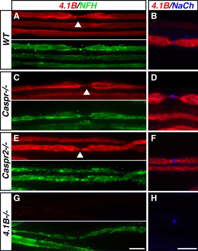
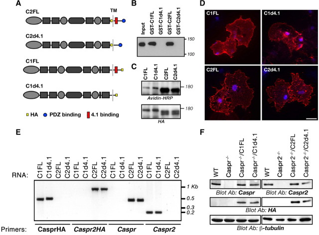
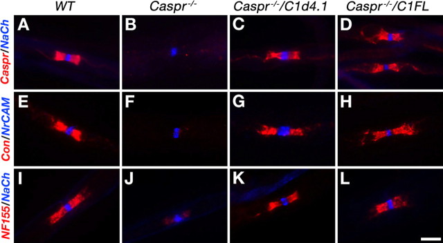
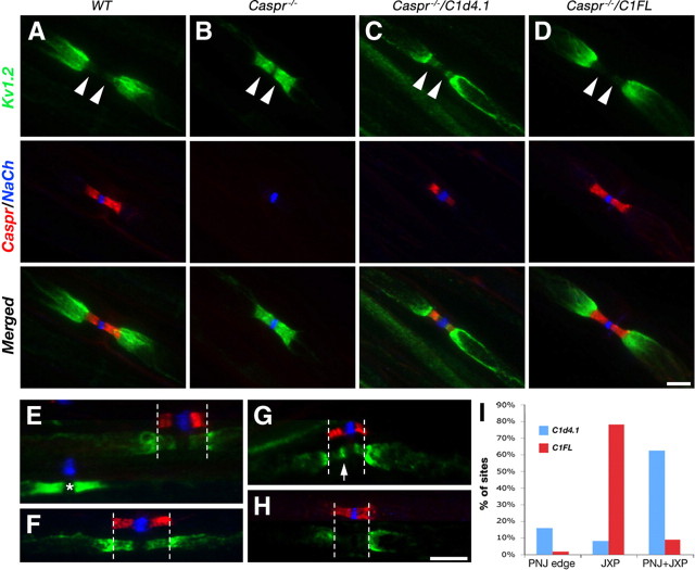
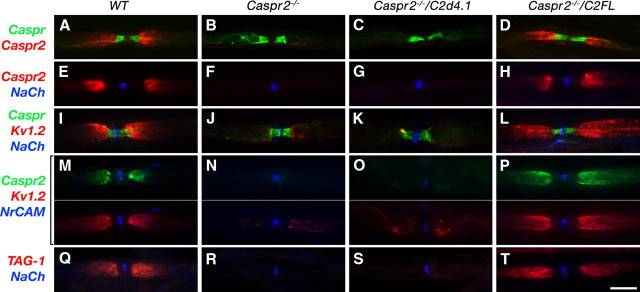
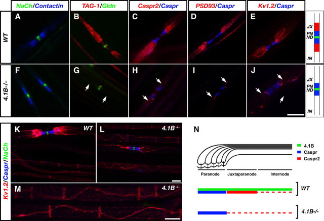
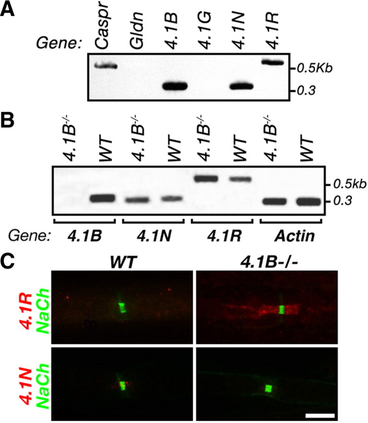
Caspr and Caspr2 regulate the formation of distinct axonal domains around the nodes of Ranvier. Caspr is required for the generation of a membrane barrier at the paranodal junction (PNJ), whereas Caspr2 serves as a membrane scaffold that clusters Kv1 channels at the juxtaparanodal region (JXP). Both Caspr and Caspr2 interact with protein 4.1B, which may link the paranodal and juxtaparanodal adhesion complexes to the axonal cytoskeleton. To determine the role of protein 4.1B in the function of Caspr proteins, we examined the ability of transgenic Caspr and Caspr2 mutants lacking their 4.1-binding sequence (d4.1) to restore Kv1 channel clustering in Caspr- and Caspr2-null mice, respectively. We found that Caspr-d4.1 was localized to the PNJ and is able to recruit the paranodal adhesion complex components contactin and NF155 to this site. Nevertheless, in axons expressing Caspr-d4.1, Kv1 channels were often detected at paranodes, suggesting that the interaction of Caspr with protein 4.1B is necessary for the generation of an efficient membrane barrier at the PNJ. We also found that the Caspr2-d4.1 transgene did not accumulate at the JXP, even though it was targeted to the axon, demonstrating that the interaction with protein 4.1B is required for the accumulation of Caspr2 and Kv1 channels at the juxtaparanodal axonal membrane. In accordance, we show that Caspr2 and Kv1 channels are not clustered at the JXP in 4.1B-null mice. Our results thus underscore the functional importance of protein 4.1B in the organization of peripheral myelinated axons.


Figures

Figure 1.
Figure 1.
Distribution of protein 4.1B in myelinated axons is not affected in the absence of Caspr or Caspr2. Immunofluorescence labeling of teased adult sciatic nerves isolated from wild-type (WT) (A, B), Caspr-null (Caspr−/−) (C, D), Caspr2-null (Caspr2−/−) (E, F), or protein 4.1B-null (4.1B−/−) (G, H) mice, using antibodies to protein 4.1B and neurofilament H (NFH) or Na+ channels (NaCh) as indicated. Images of the individual green and red channels are shown in A, C, E, and G. The arrowheads indicate the location of the nodes. Protein 4.1B is found at the PNJ, JXP, and internodes in the presence or absence of Caspr or Caspr2. Scale bars, 10 μm.
Figure 2.
Figure 2.
Characterization of Caspr and Caspr2 transgenes. A, Schematic representation of the different constructs used. The cytoplasmic domain of Caspr contains a 4.1-binding sequence (red square), whereas that of Caspr2 contains both 4.1 and PDZ (blue circle) domain binding sequences. C1FL and C2FL encode a full-length HA-tagged (yellow square) Caspr and Caspr2, respectively. The C1d4.1 and C2d4.1 constructs encode for Caspr and Caspr2 lacking their protein 4.1-binding region. B, Interaction with protein 4.1B. GST fusion proteins containing the cytoplasmic domain of Caspr (C1CT) or Caspr2 (C2CT), or cytoplasmic domains lacking their putative protein 4.1-binding sequence (C1d4.1 or C2d4.1) used to pull down myc-tagged protein 4.1B from lysates of 4.1B-transfected HEK293 cells. Protein complexes were subjected to immunoblot analysis using an antibody to myc tag. A sample of the cell lysate (Input) was used as control for protein 4.1B expression. The location of molecular mass markers is shown on the right in kilodaltons. C, D, Caspr2 transgenes reach the cell surface. C, Cell surface biotinylation was performed using HEK293 cells expressing Caspr and Caspr2 transgenes. Cell lysates were subjected to immunoprecipitation with an antibody to HA tag followed by immunoblotting with the same antibody (bottom panel) or with streptavidin-HRP (top panel). The location of molecular mass markers is shown on the right in kilodaltons. D, Nonpermeabilized COS-7 cells expressing Caspr transgenes and contactin, or Caspr2 transgenes, were labeled with an antibody to the extracellular domain of Caspr (top panels) or Caspr2 (bottom panels). Cell nuclei are labeled with DAPI (4′,6′-diamidino-2-phenylindole) (blue). Scale bar, 30 μm. E, Expression of the transgenic transcripts in peripheral sensory neurons. Dorsal root ganglion mRNA isolated from Caspr−/−/C1FL (C1FL), Caspr−/−/C1d4.1 (C1d4.1), Caspr2−/−/C2FL (C2FL), and Caspr2−/−/C2d4.1 (C2d4.1) transgenic mice were used as a template for RT-PCR using specific primer sets that recognize each of the endogenous genes (Caspr and Caspr2), or each of the transgenes (CasprHA and Caspr2HA). The location of size markers is shown on the right in kilobases. F, All transgenic proteins are targeted to the axon. Immunoblot analysis of sciatic nerves isolated from wild-type (WT), Caspr or Caspr2 nulls, or the four transgenic mice, using antibodies to Caspr, Caspr2, HA tag, or β-tubulin as indicated.
Figure 3.
Figure 3.
Caspr transgenes are localized at the PNJ and rescued the paranodal adhesion complex. Double immunofluorescence labeling of teased sciatic nerves isolated from wild-type mice (WT), Caspr nulls (Caspr−/−), or Caspr nulls expressing the C1d4.1 (Caspr−/−/C1d4.1), or the FL (Caspr−/−/C1FL) transgenes, using antibodies to Caspr and Na+ channels (NaCh) (A–D), contactin (Con) and NrCAM (E–H), or NF155 and Na+ channels (I–L). Scale bar, 5 μm.
Figure 4.
Figure 4.
Kv1 channels are not efficiently excluded from the PNJ in Caspr−/−/C1d4.1 mutant mice. Triple immunofluorescence labeling of teased sciatic nerves isolated from wild-type mice (WT) (A), Caspr nulls (Caspr−/−) (B), or Caspr nulls expressing the C1FL (Caspr−/−/FL) (C) or the C1d4.1 (Caspr−/−/C1d4.1) (D–H) transgenes, using antibodies to Caspr (red), Kv1.2 (green), and Na+ channels (NaCh; blue). The arrowheads in A–D mark the location of the PNJ. Staining for Caspr and NaCh was shifted in E–H to reveal Kv1.2 immunoreactivity. Note that, whereas the expression of the C1FL transgene resulted in complete exclusion of Kv1.2 to the JXP (D), these channels were present to various degrees at the PNJ in Caspr−/−/C1d4.1 axons (D–G). The asterisk in E marks a nodal site that does not express the transgene. An arrow in G points to the accumulation of Kv1.2 in a line between the nodes and the PNJ. I, Quantitation of Kv1.2 distribution in sciatic nerves of Caspr−/−/C1d4.1 or Caspr−/−/C1FL transgenes. Percentage of sites along sciatic nerves in which Kv1.2 channels are located at the juxtaparanodes (JXP), at both juxtaparanodes and paranodes (JXP+PNJ), or are concentrated at the edges of the PNJ (PNJ edge). Scale bars, 10 μm.
Figure 5.
Figure 5.
Localization of Caspr2 at the JXP requires its protein 4.1-binding site. Immunofluorescence labeling of teased sciatic nerves isolated from wild-type mice (WT) (A, E, I, M), Caspr2 nulls (Caspr2−/−) (B, F, J, N), or Caspr2 nulls expressing the C2d4.1 (Caspr2−/−/C2d4.1) (C, G, K, O) or the C2FL (Caspr2−/−/C2FL) (D, H, L, P) transgenes, using different antibody combinations as indicated on the left. Merged images are shown in A–L and Q–T, whereas in M–P the red and green channels are shown in separate panels. In contrast to the C2FL transgene, C2d4.1, Kv1 channels, and TAG-1 are not clustered at the JXP. Scale bar, 10 μm.
Figure 6.
Figure 6.
Impaired formation of the JXP domain in peripheral nerves of mice lacking protein 4.1B. Teased sciatic nerves isolated from wild-type mice (WT) (A–E), or 4.1B-null mice (4.1B−/−) (F–J), were labeled with antibodies to Na+ channels (NaCh) and contactin (A, F), TAG-1 and gliomedin (Gldn) (B, G), Caspr2 and Caspr (C, H), PSD93 and Caspr (D, I), or Kv1.2 and Caspr (E, J). The organization of the nodal environs in WT and 4.1B−/− nerves is schematically depicted on the right. JX, Juxtaparanodes; PN, paranodes; ND, nodes of Ranvier; IN, internodes. K–M, Triple immunofluorescence labeling of teased sciatic nerve fibers isolated from wild-type (K) or 4.1B−/− (L, M) animals, using antibodies to Kv1.2, Caspr, and NaCh. Note that Kv1.2 channels are located in a double line along the juxtamesaxon and below the Schmidt–Lanterman incisures. Scale bars, 10 μm. N, Schematic drawing illustrating the localization of protein 4.1B, Caspr, and Caspr2 along myelinated axons in wild-type and 4.1B−/− mice.
Figure 7.
Figure 7.
Protein 4.1R is concentrated at the PNJ in the absence of 4.1B. A, Expression of protein 4.1 family members in peripheral sensory neurons. Purified rat DRG neuron mRNA was used as a template for RT-PCR using specific primer sets that recognize each of the 4.1 genes. Caspr and gliomedin (Gldn) were used as controls for genes that are expressed in DRG neurons or Schwann cells, respectively. The location of size markers is shown on the right in kilobases. B, Expression of neuronal 4.1 genes in the absence of protein 4.1B. RT-PCR analysis was done on DRG mRNA isolated from 4.1B−/− and wild-type mice using specific primers that recognize 4.1B, 4.1N, 4.1R, and actin genes. C, Protein 4.1R is concentrated at the PNJ in 4.1B−/−. Double immunofluorescence labeling of teased sciatic nerves isolated from wild-type mice (WT), or protein 4.1B nulls (4.1B−/−), using antibodies to Na+ channels (NaCh), and protein 4.1R or protein 4.1N as indicated. Scale bar, 10 μm.

Similar articles

Cited by

References

    1. Arroyo EJ, Xu YT, Zhou L, Messing A, Peles E, Chiu SY, Scherer SS. Myelinating Schwann cells determine the internodal localization of Kv1.1, Kv1.2, Kvbeta2, and Caspr. J Neurocytol. 1999;28:333–347. - PubMed
    1. Arroyo EJ, Sirkowski EE, Chitale R, Scherer SS. Acute demyelination disrupts the molecular organization of peripheral nervous system nodes. J Comp Neurol. 2004;479:424–434. - PubMed
    1. Baumgartner S, Littleton JT, Broadie K, Bhat MA, Harbecke R, Lengyel JA, Chiquet-Ehrismann R, Prokop A, Bellen HJ. A Drosophila neurexin is required for septate junction and blood-nerve barrier formation and function. Cell. 1996;87:1059–1068. - PubMed
    1. Beekman JM, Bakema JE, van der Poel CE, van der Linden JA, van de Winkel JG, Leusen JH. Protein 4.1G binds to a unique motif within the Fc gamma RI cytoplasmic tail. Mol Immunol. 2008;45:2069–2075. - PubMed
    1. Bennett V, Baines AJ. Spectrin and ankyrin-based pathways: metazoan inventions for integrating cells into tissues. Physiol Rev. 2001;81:1353–1392. - PubMed

Publication types

MeSH terms