. 2010 Feb 17;30(7):2496-503.
doi: 10.1523/JNEUROSCI.6254-09.2010.

Deletion of GRK1 causes retina degeneration through a transducin-independent mechanism

Affiliations

Deletion of GRK1 causes retina degeneration through a transducin-independent mechanism

Jie Fan et al. J Neurosci. .

Abstract

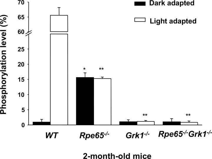
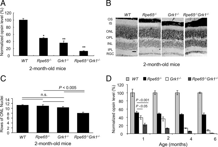
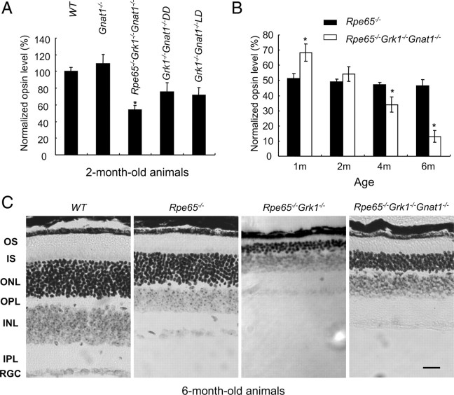
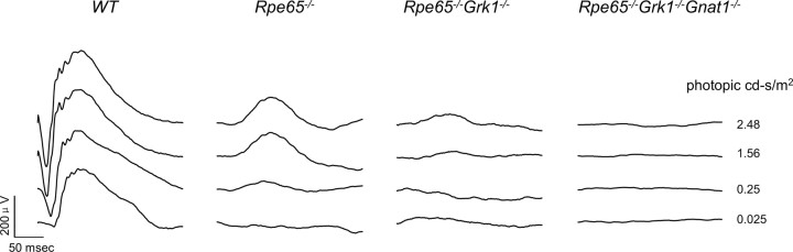
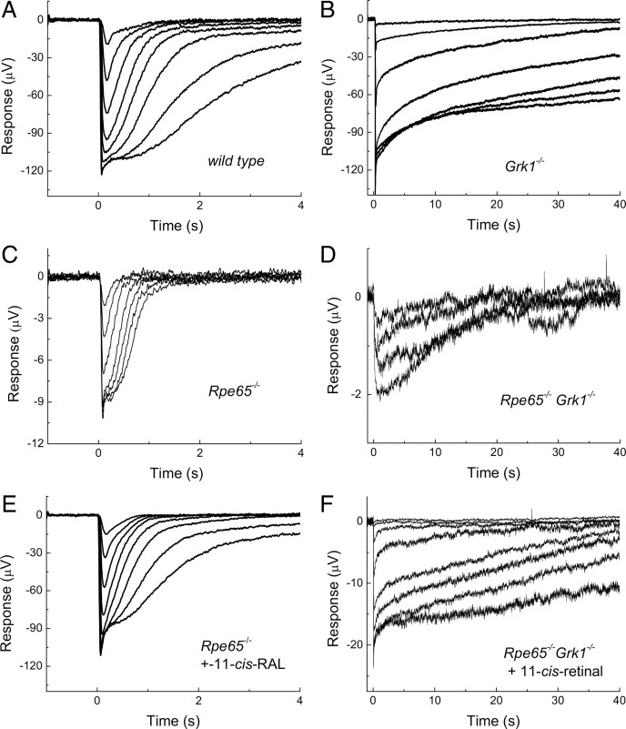
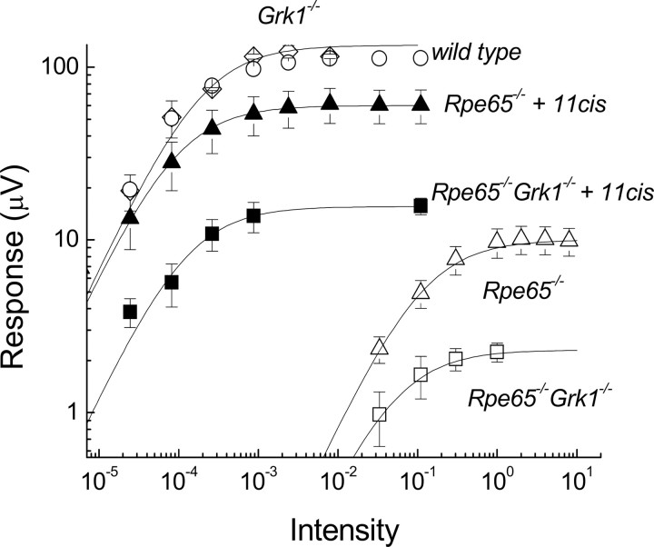
Rpe65(-/-) mice are unable to produce 11-cis-retinal, the chromophore of visual pigments. Consequently, the pigment is present as the apoprotein opsin with a minute level of pigment containing 9-cis-retinal as chromophore. Notably, a 10-20% fraction of this opsin is mono-phosphorylated independently of light conditions. To determine the role of rhodopsin kinase (GRK1) in phosphorylating this opsin and to test whether eliminating this phosphorylation would accelerate photoreceptor degeneration, we generated the Rpe65(-/-)Grk1(-/-) mouse. The retinae of Rpe65(-/-)Grk1(-/-) mice had negligible opsin phosphorylation, extensive degeneration with decreased opsin levels, and diminished light-evoked rod responses relative to Rpe65(-/-) mice. These data show that opsin phosphorylation in the Rpe65(-/-) mouse is due to the action of GRK1 and is neuroprotective. However, despite the higher activity of unphosphorylated opsin, the severe loss of opsin in the rapidly degenerating Rpe65(-/-)Grk1(-/-) mice resulted in lower overall opsin activity and in higher rod sensitivity compared with Rpe65(-/-) mice. In Rpe65(-/-)Grk1(-/-)Gnat1(-/-) mice where transduction activation was blocked, degeneration was only partially prevented. Therefore, increased opsin activity in the absence of phosphorylation was not the only mechanism for the accelerated retinal degeneration. Finally, the deletion of GRK1 triggered retinal degeneration in Grk1(-/-) mice after 1 month, even in the absence of apo-opsin. This degeneration was independent of light conditions and occurred even in the absence of transducin in Grk1(-/-)Gnat1(-/-) mice. Taken together, our results demonstrate a light-independent mechanism for retinal degeneration in the absence of GRK1, suggesting a second, not previously recognized role for that kinase.


Figures

Figure 1.
Figure 1.
Effect of deletion of Rpe65 and Grk1 on opsin phosphorylation. Retinae of cyclic-light-reared, 2-month-old WT, Rpe65−/−, Grk1−/−, and Rpe65−/−Grk1−/− mice were homogenized in 8 m urea and digested with endoproteinase Asp-N in 10 mm Tris buffer at pH 7.6 to cleave the opsin C terminus, which was analyzed online with an LCQ mass spectrometer. In the Grk1−/− and Rpe65−/−Grk1−/− mice, no significant opsin phosphorylation was observed. White bars, Animals exposed to room light for 6 h; black bars, animals dark-adapted for 12 h. Data are shown as the percentage of rhodopsin C terminus containing phosphorylation, independent of the multiplicity of phosphorylation, and presented as mean ± SEM; n = 3.
Figure 2.
Figure 2.
Effect of deletion of Rpe65 and Grk1on retinal opsin levels and morphology. A, Opsin levels were calculated from rhodopsin that formed upon the addition of 11-cis-retinal. Data were generated from 2-month-old cyclic-light-reared mice. The relative opsin levels in each strain were normalized to the average of wild-type opsin concentration and shown as a fraction of the wild type. B, Retinal morphology of 2-month-old WT, Rpe65−/−, Grk1−/−, and Rpe65−/−Grk1−/− mice. Light micrographs of paraffin-embedded retinae sections from the superior central region of the retina. While morphology of the inner retina was unaffected by the different genotypes at that resolution, the outer retina showed shortening of the inner and outer segments progressing in severity from the Rpe65−/− < Grk1−/−Rpe65−/−Grk1−/− (scale bar, 30 μm). INL, Inner nuclear layer; IPL, inner plexiform layer; IS, inner segments; OPL, outer plexiform layer; OS, outer segments; RGC, retinal ganglion cells; n.s., not significant. C, Bar graph representing rows of photoreceptor cell nuclei counted in the superior central region of the retinae of 2-month-old mice from the 4 strains; mean ± SEM; n = 5. The cell loss was significant for the Rpe65−/−Grk1−/− mice. D, Opsin levels with age, assayed as for A. The relative opsin levels in each strain were normalized to the average of wild-type opsin concentration at each age, and shown as the fraction of the wild type. Gray bar, WT mice; black bar, Rpe65−/− mice; white bar, Grk1−/− mice; hatched bar, Rpe65−/−Grk1−/− mice.
Figure 3.
Figure 3.
Effect of deletion of Gnat1 on photoreceptor degeneration in the absence of Grk1. A, Opsin levels were calculated as for Figure 2A. Data are for 2-month-old mice. The relative opsin levels were normalized to the average of wild-type opsin concentration and shown as the fraction of wild type. B, Opsin levels from Rpe65−/−Grk1−/−Gnat1−/− mice with age, assayed as for A. C, Retinal morphology in 6-month-old WT, Rpe65−/−, Rpe65−/−Grk1−/− and Rpe65−/−Grk1−/−Gnat1−/− mice. Light micrographs of paraffin-embedded retinae of 6-month-old cyclic-light-reared animals were taken from the superior central region of the eye to compare the thicknesses of the different layers. Note the extreme degeneration in the Rpe65−/−Grk1−/− retina. Scale bar, 20 μm. INL, Inner nuclear layer; IPL, inner plexiform layer; IS, inner segments; OPL, outer plexiform layer; OS, outer segments; RGC, retinal ganglion cells.
Figure 4.
Figure 4.
Scotopic transcorneal ERG responses. Families of single-flash ERGs were recorded from 2-month-old WT, Rpe65−/−, and Rpe65−/−Grk1−/− mice in response to increasing light intensities 2.48 × 10⁻², 2.48 × 10⁻¹, 1.56 and 2.48 cd*s/m2.
Figure 5.
Figure 5.
A–F, Response families of transretinal ERG recordings from WT (A), Grk1−/− (B), Rpe65−/− (C, E), and Rpe65−/−Grk1−/− (D, F) mice. Responses in E and F were recorded after application of 11-cis-retinal. Flashes of white light were delivered at time 0 in 0.5 log incremental steps except for the last three traces in C, which were delivered in twofold increments by doubling the flash duration. The dimmest flashes were delivered at −1.5 log (C, D), and −4.5 log (A, B, E, and F) unit attenuation.
Figure 6.
Figure 6.
Amplitude of transretinal ERG a-wave as a function of flash intensity in WT (○; n = 3), Grk1−/− (□ n = 3), Rpe65−/− (Δ; n = 7), Rpe65−/−Grk1−/− (□; n = 9), Rpe65−/− with 11-cis-retinal (▲; n = 9), and Rpe65−/−Grk1−/− with 11-cis-retinal (■; n = 6) retinae. The intensity-response data are fitted by Equation 1. Data from Rpe65−/− Grk1−/− mice are from animals raised in cyclic-light conditions. Data are presented as mean ± SEM (n = 8).

Similar articles

Cited by

References

    1. Ablonczy Z, Knapp DR, Darrow R, Organisciak DT, Crouch RK. Mass spectrometric analysis of rhodopsin from light damaged rats. Mol Vis. 2000;6:109–115. - PubMed
    1. Ablonczy Z, Crouch RK, Goletz PW, Redmond TM, Knapp DR, Ma JX, Rohrer B. 11-cis-retinal reduces constitutive opsin phosphorylation and improves quantum catch in retinoid-deficient mouse rod photoreceptors. J Biol Chem. 2002;277:40491–40498. - PubMed
    1. Adams RA, Liu X, Williams DS, Newton AC. Differential spatial and temporal phosphorylation of the visual receptor, rhodopsin, at two primary phosphorylation sites in mice exposed to light. Biochem J. 2003;374:537–543. - PMC - PubMed
    1. Arshavsky VY. Rhodopsin phosphorylation: from terminating single photon responses to photoreceptor dark adaptation. Trends Neurosci. 2002;25:124–126. - PubMed
    1. Batten ML, Imanishi Y, Maeda T, Tu DC, Moise AR, Bronson D, Possin D, Van Gelder RN, Baehr W, Palczewski K. Lecithin-retinol acyltransferase is essential for accumulation of all-trans-retinyl esters in the eye and in the liver. J Biol Chem. 2004;279:10422–10432. - PMC - PubMed

Publication types

MeSH terms

LinkOut - more resources