. 2010 Feb 17;30(7):2533-41.
doi: 10.1523/JNEUROSCI.5768-09.2010.

Brain-derived neurotrophic factor regulates hedonic feeding by acting on the mesolimbic dopamine system

Affiliations

Brain-derived neurotrophic factor regulates hedonic feeding by acting on the mesolimbic dopamine system

Joshua W Cordeira et al. J Neurosci. .

Abstract

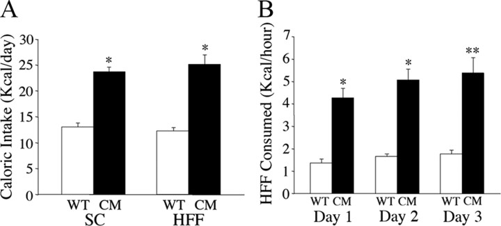
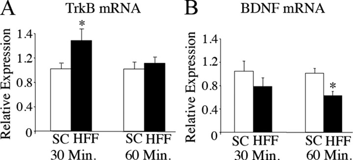
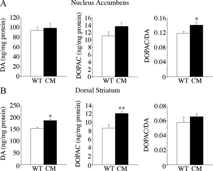
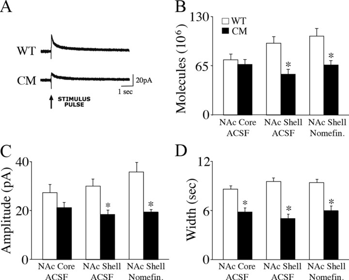
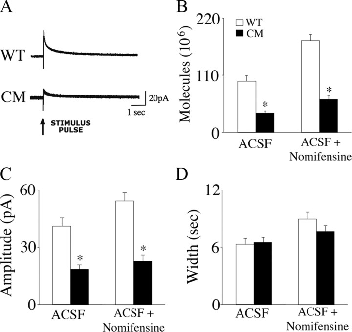
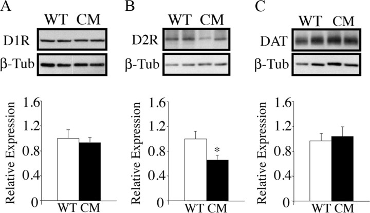
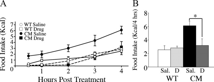
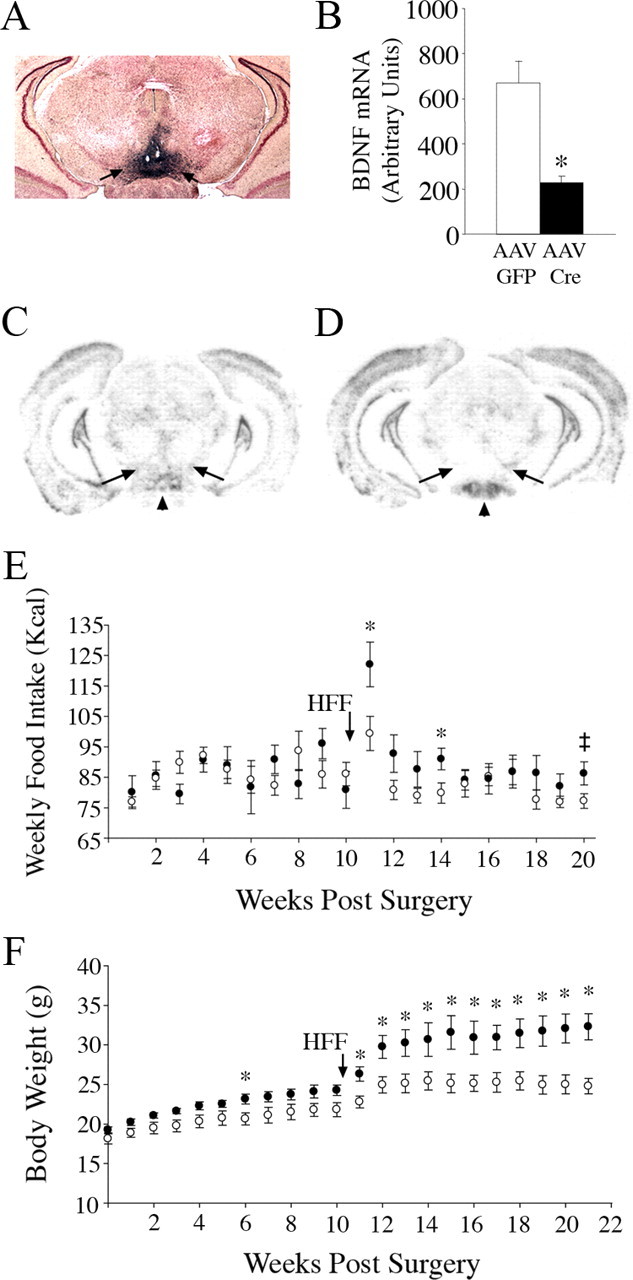
Brain-derived neurotrophic factor (BDNF) and its receptor, TrkB, play prominent roles in food intake regulation through central mechanisms. However, the neural circuits underlying their anorexigenic effects remain largely unknown. We showed previously that selective BDNF depletion in the ventromedial hypothalamus (VMH) of mice resulted in hyperphagic behavior and obesity. Here, we sought to ascertain whether its regulatory effects involved the mesolimbic dopamine system, which mediates motivated and reward-seeking behaviors including consumption of palatable food. We found that expression of BDNF and TrkB mRNA in the ventral tegmental area (VTA) of wild-type mice was influenced by consumption of palatable, high-fat food (HFF). Moreover, amperometric recordings in brain slices of mice depleted of central BDNF uncovered marked deficits in evoked release of dopamine in the nucleus accumbens (NAc) shell and dorsal striatum but normal secretion in the NAc core. Mutant mice also exhibited dramatic increases in HFF consumption, which were exacerbated when access to HFF was restricted. However, mutants displayed enhanced responses to D(1) receptor agonist administration, which normalized their intake of HFF in a 4 h food intake test. Finally, in contrast to deletion of Bdnf in the VMH of mice, which resulted in increased intake of standard chow, BDNF depletion in the VTA elicited excessive intake of HFF but not of standard chow and increased body weights under HFF conditions. Our findings indicate that the effects of BDNF on eating behavior are neural substrate-dependent and that BDNF influences hedonic feeding via positive modulation of the mesolimbic dopamine system.


Figures

Figure 1.
Figure 1.
BDNF2L/2LCk-cre mutant mice exhibit increased intake of palatable high-fat food. A, Caloric intake of wild-type (WT) and BDNF2L/2LCk-cre conditional mutant (CM) mice that were fed SC or palatable HFF ad libitum (n = 10; *p < 0.0001). B, Caloric intake of high-fat chow of wild-type and BDNF2L/2LCk-cre mutant mice during a 1 h restricted period over 3 consecutive days (n = 10; *p < 0.0001; **p = 0.0001).
Figure 2.
Figure 2.
Intake of palatable high-fat food alters levels of BDNF and TrkB mRNA in the VTA of wild-type mice. A, Expression levels of TrkB mRNA in the ventral tegmental area of wild-type mice fed SC or HFF chow for 30 or 60 min as measured by quantitative RT-PCR analysis (n = 5; *p = 0.04). B, Relative expression of BDNF mRNA in the ventral tegmental area of wild-type mice fed standard or high-fat chow for 30 or 60 min (n = 5; *p = 0.004). Data are expressed as fold difference of HFF-fed relative to SC-fed wild-type animals. The mean value for SC-fed mice was set at 1. p values were calculated based on 2{Δ}{Δ}Ct values for each sample.
Figure 3.
Figure 3.
Dopamine content in nucleus accumbens and dorsal striatum of BDNF2L/2LCk-cre mutant mice. A, HPLC measurements of DA and DOPAC content and DOPAC/DA ratios in the nucleus accumbens of wild-type control (WT) and BDNF2L/2LCk-cre conditional mutant (CM) mice (n = 5; *p = 0.02). B, Dopamine and DOPAC content and DOPAC/DA ratios in the dorsal striatum of wild-type control and BDNF2L/2LCk-cre conditional mutant mice (n = 5; *p = 0.002; **p = 0.007).
Figure 4.
Figure 4.
Evoked release of dopamine in the nucleus accumbens shell is impaired in BDNF2L/2LCk-cre conditional mutant mice. A, Representative amperometric traces of electrical stimulation-evoked dopamine release in acute coronal accumbens slices of wild-type (WT) and BDNF2L/2LCk-cre conditional mutant (CM) mice. Stimulation electrodes and carbon fiber recording microelectrodes were positioned in the shell region of the nucleus accumbens, which receives the majority of the dopaminergic projections from the VTA. Data were acquired at 50 kHz and digitally postfiltered at 1 kHz. Arrow points to onset of electrical single pulse (2 ms of 0.5 mA). B, Mean evoked dopamine molecules in the nucleus accumbens core (NAc Core) and shell (NAc Shell) in brain slices from wild-type and BDNF2L/2LCk-cre conditional mutant mice in ACSF and ACSF with the dopamine reuptake inhibitor nomifensine (Nomifen.). *p < 0.01. C, Evoked dopamine signal amplitude in the NAc core and shell of wild-type and BDNF2L/2LCk-cre mutant mice. *p < 0.01. D, Evoked dopamine signal width in the NAc core and shell of wild-type and BDNF2L/2LCk-cre mutant mice. *p < 0.01.
Figure 5.
Figure 5.
BDNF2L/2LCk-cre conditional mutants exhibit deficits in evoked release of dopamine in the dorsal striatum. *p < 0.01. A, Representative amperometric traces of electrical stimulation-evoked dopamine release in acute coronal striatal slices from wild-type (WT) and BDNF2L/2LCk-cre conditional mutant (CM) mice. Stimulation electrodes and carbon fiber recording microelectrodes were positioned in the medial–dorsal region of the striatum. Data were acquired at 50 kHz and digitally postfiltered at 1 kHz. The arrow points to onset of electrical single pulse (2 ms of 0.5 mA). B, Mean evoked dopamine molecules in the dorsal striatum of wild-type and BDNF2L/2LCk-cre conditional mutant mice in ACSF and ACSF with the dopamine reuptake inhibitor nomifensine. *p < 0.01. C, Evoked dopamine signal amplitude in the dorsal striatum of wild-type and BDNF2L/2LCk-cre mutant mice. *p < 0.01. D, Evoked dopamine signal width in the dorsal striatum of wild-type and BDNF2L/2LCk-cre mutant mice. *p < 0.01.
Figure 6.
Figure 6.
Expression of D2 receptor protein is reduced in the dorsal striatum of BDNF2L/2LCk-cre conditional mutants. A, Representative Western blot and densitometry analysis measuring D1R protein content in the dorsal striatum of wild-type (WT) and BDNF2L/2LCk-cre conditional mutant (CM) mice (n = 5). Data are expressed as expression of BDNF mutant mice relative to wild-type controls. B, Expression of D2R protein in the dorsal striatum of wild-type and BDNF conditional mutant mice (n = 5; p = 0.03). C, Expression of DAT in the dorsal striatum of wild-type and BDNF conditional mutant mice (n = 5).
Figure 7.
Figure 7.
Peripheral administration of the D1 receptor selective agonist SKF38393 reduced consumption of palatable high-fat food of BDNF2L/2LCk-cre mutant mice. A, Time course for HFF caloric intake of fed wild-type (WT) and BDNF2L/2LCk-cre conditional mutant (CM) mice following peripheral administration of saline vehicle or SKF38393 drug (n = 5 per group). B, Four-hour cumulative HFF intake of fed wild-type and BDNF mutant mice following saline vehicle (Sal.) or SKF38393 drug (D) treatment. *p = 0.04, n = 5 per group.
Figure 8.
Figure 8.
Selective targeting of Bdnf in the VTA results in increased intake of palatable HFF and higher body weights. A, Representative coronal brain section from a Rosa-β-gal reporter mouse that was delivered AAV2/1-Cre to the VTA. B, Densitometry of BDNF mRNA signal detected by in situ hybridization analysis in AAV2/1-GFP (n = 6) and AAV2/1-Cre (n = 5) injected mice. *p = 0.004. C, D, In situ hybridization analysis of BDNF mRNA expression in coronal brain sections from floxed Bdnf mice injected with AAV2/1-GFP (C) or AAV2/1-Cre (D) in the VTA. Arrows indicate the VTA and arrowhead indicates the mammillary nucleus. E, Weekly caloric intake of AAV2/1-GFP (open circles, n = 6) and AAV2/1-Cre (closed circles, n = 5) injected mice under standard and high-fat chow conditions (*p < 0.05; ‡, p = 0.06). Arrow indicates time point when mice were switched from a standard to a high-fat chow diet. F, Body weights of AAV2/1-GFP (open circles, n = 6) and AAV2/1-Cre (closed circles, n = 5) injected mice over 21 weeks following stereotaxic surgery and under standard and high-fat chow conditions (*p < 0.05). Arrow indicates time point when mice were switched from a standard to a high-fat chow diet.

Similar articles

Cited by

References

    1. Abizaid A, Liu ZW, Andrews ZB, Shanabrough M, Borok E, Elsworth JD, Roth RH, Sleeman MW, Picciotto MR, Tschöp MH, Gao XB, Horvath TL. Ghrelin modulates the activity and synaptic input organization of midbrain dopamine neurons while promoting appetite. J Clin Invest. 2006;116:3229–3239. - PMC - PubMed
    1. Baldo BA, Alsene KM, Negron A, Kelley AE. Hyperphagia induced by GABAA receptor-mediated inhibition of the nucleus accumbens shell: dependence on intact neural output from the central amygdaloid region. Behav Neurosci. 2005;119:1195–1206. - PubMed
    1. Baquet ZC, Bickford PC, Jones KR. Brain-derived neurotrophic factor is required for the establishment of the proper number of dopaminergic neurons in the substantia nigra pars compacta. J Neurosci. 2005;25:6251–6259. - PMC - PubMed
    1. Bariohay B, Lebrun B, Moyse E, Jean A. Brain-derived neurotrophic factor plays a role as an anorexigenic factor in the dorsal vagal complex. Endocrinology. 2005;146:5612–5620. - PubMed
    1. Bassareo V, Di Chiara G. Differential influence of associative and nonassociative learning mechanisms on the responsiveness of prefrontal and accumbal dopamine transmission to food stimuli in rats fed ad libitum. J Neurosci. 1997;17:851–861. - PMC - PubMed

Publication types

MeSH terms