. 2010 Feb 17;30(7):2600-10.
doi: 10.1523/JNEUROSCI.3744-09.2010.

Cilia organize ependymal planar polarity

Affiliations

Cilia organize ependymal planar polarity

Zaman Mirzadeh et al. J Neurosci. .

Abstract

Multiciliated epithelial cells, called ependymal cells, line the ventricles in the adult brain. Most ependymal cells are born prenatally and are derived from radial glia. Ependymal cells have a remarkable planar polarization that determines orientation of ciliary beating and propulsion of CSF. Disruption of ependymal ciliary beating, by injury or disease, results in aberrant CSF circulation and hydrocephalus, a common disorder of the CNS. Very little is known about the mechanisms guiding ependymal planar polarity and whether this organization is acquired during ependymal cell development or is already present in radial glia. Here we show that basal bodies in ependymal cells in the lateral ventricle walls of adult mice are polarized in two ways: (1) rotational; angle of individual basal bodies with respect to their long axis and (2) translational; the position of basal bodies on the apical surface of the cell. Conditional ablation of motile cilia disrupted rotational orientation, but translational polarity was largely preserved. In contrast, translational polarity was dramatically affected when radial glial primary cilia were ablated earlier in development. Remarkably, radial glia in the embryo have a translational polarity that predicts the orientation of mature ependymal cells. These results suggest that ependymal planar cell polarity is a multistep process initially organized by primary cilia in radial glia and then refined by motile cilia in ependymal cells.


Figures

Figure 1.
Figure 1.
Functional planar polarity of the ependyma. Diagram at top right shows the lateral wall of the lateral ventricle divided by 4 lines into the regions analyzed: AD (anterior–dorsal), AV (anterior–ventral), and PM (posterior–middle). Line (a1) bisects the adhesion area into an anterior and posterior half. Line (a2) bisects the region between line (a1) and the anterior boundary of the wholemount. Line (b1) and (b2) run parallel along the anterior–posterior axis of the wholemount and divide the dorsal-ventral axis into 3 equal rows. A, Top is from 89 consecutive frames of Movie S1 that were merged into a single picture. Fluorescent microbeads were deposited (*) on the surface of a wholemount of the lateral wall of the lateral ventricle. Oriented flow lines reveal planar polarized movement of beads above the ventricular wall surface driven by ependymal cilia. Each flow line represents sequential positions of a single bead. Yellow arrows indicate direction of flow. Anterior (a) and dorsal (d) directions are shown by white arrows. The adhesion area is where the medial and lateral walls are fused within the ventricular cavity; it is not covered by ependyma. Red boxes outline flow in regions AD, AV, and PM where cellular analyses in Figure 2 and supplemental Figure S2 (available at www.jneurosci.org as supplemental material) were performed. These regions are readily identified with respect to the adhesion area and their flow differs significantly: anteroventral in AD and PM, posteroventral in AV. Bottom shows individual frames of this movie. Scale bar, 0.5 mm. B, High-power image of 2 ependymal cell basal bodies demonstrating the ultrastructure of the barrel and the basal foot (arrowheads) in cross-section. Scale bar, 200 nm. C, En-face electron micrograph near the apical surface of 2 adjacent ependymal cells in region AD. The orientation of this image and all subsequent images in this study correspond to the orientation of the wholemount in A. Note clustering of basal bodies to one side of the apical surface in both cells. Higher-power images of the basal bodies are shown in D and E (boxed regions). Scale bar, 2 μm. D, E, Some basal bodies in C were transected at the level of the basal foot. Orientation of the basal foot (blue arrows) is consistent within each cell and between cells and points in the direction of CSF flow (black arrow in (C). Scale bar, 0.25 μm.
Figure 2.
Figure 2.
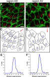
Basal body patch position is an anatomical indicator of ependymal planar polarity. A, B, Confocal images from regions AD and AV (see Fig. 1A) of lateral wall wholemounts stained for β-catenin (green) and γ-tubulin (red). In both regions, the patch of basal bodies is located on the “downstream” side of the apical surface with respect to CSF flow (compare with flow in boxed regions in Fig. 1A). Scale bar, 10 μm. C, D, Traces of the apical surfaces and basal body patches in A, B. Basal body patch position was measured relative to the apical surface using a vector connecting the centers of these two traced regions for each cell. The angle of this vector was called the basal body (BB) patch angle. The red arrows indicate the direction of the median angle in the region. E, F, For each high-power field analyzed, the difference between each cell's BB patch angle and the median BB patch angle in the field was calculated and these data were plotted on a histogram. The narrow distribution around 0° revealed that BB patch angles in these regions were highly oriented.
Figure 3.
Figure 3.
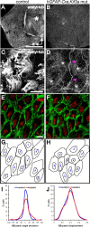
Planar polarity of basal body patches does not require motile (9 + 2) cilia. A, B, Low-power confocal images of lateral wall wholemounts from the LV of hGFAP::Cre;Kif3afl/fl mutants and control littermates stained for acetylated-tubulin. Tufts of cilia, seen as small white puncta on the ependymal surface of controls at this magnification, are missing in mutants. Asterisk on the control wholemount indicates the adhesion area, which is not present in the mutant, likely due to hydrocephalus. Anterior (a) and dorsal (d) directions are indicated by arrows. Scale bar, 0.5 mm. C, D, High-power confocal images from the wholemounts in A, B show that while microtubule-based cilia were absent on the apical surface of mutant ependymal cells, the microtubule network (*) within these cells was preserved. Note also that intraventricular axons (magenta arrowheads), stained by acetylated-tubulin, were seen on the ventricular surface of both wholemounts, although more clearly on the cilia-deficient mutant ependyma. Intraventricular axons have been described previously (Vígh et al., 2004). Scale bar, 10 μm. E, F, Confocal images of control and hGFAP::Cre;Kif3afl/fl mutant lateral wall wholemounts stained for β-catenin (green) and γ-tubulin (red). Basal bodies in mutant ependymal cells were maintained in tightly clustered patches. The planar polarized position of these patches was also largely preserved in the absence of motile cilia. Scale bar, 10 μm. G, H, Traces of the apical surfaces and basal body patches in E, F, with vectors indicating the relative position of the BB patch. I, Histogram showing the distribution of BB patch angles around the median in control (blue) and mutant (red) ependyma. As indicated by the narrow distribution, the mutant ependyma had highly oriented BB patch angles (n = 5 mutants (790 cells), 5 control littermates (624 cells); curve fit test, p < 0.0001). J, Histogram showing the distribution of normalized BB patch displacement from center in control (blue) and mutant (red) ependyma (curve fit test, p = 0.9839).
Figure 4.
Figure 4.
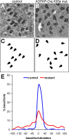
Alignment of basal body rotational orientation requires motile cilia. A, B, En-face electron micrographs from the apical surface of control and hGFAP::Cre;Kif3afl/fl mutant ependymal cells demonstrated the organization and rotational orientation of their basal bodies. C, D, Schematic drawings of the basal bodies shown in A, B illustrate the misalignment of basal body rotational orientation, indicated by triangle-shaped basal feet, in hGFAP::Cre;Kif3afl/fl mutants. E, Histograms showing the distribution of basal foot angles around the median in control and hGFAP::Cre;Kif3afl/fl mutant ependyma demonstrated that basal body rotational orientation is significantly reduced in the absence of motile cilia (p < 0.0001, n = 3 control mice with 330 basal bodies in 65 cells analyzed, 3 mutant mice with 750 basal bodies in 92 cells).
Figure 5.
Figure 5.
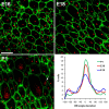
Planar polarity of basal body position in radial glia. Confocal images of lateral wall wholemounts from E16, E18, and P1 wild-type mice stained for β-catenin (green) and γ-tubulin (red). Images were taken from a mid-ventral region of the wholemount near the foramen of Monro (supplemental Fig. S9, available at www.jneurosci.org as supplemental material, arrowhead). At successively older ages, radial glial apical surfaces expanded and by P1, some radial glia had already transformed into ependymal cells with multiple basal bodies. Note that some cells had dense punctate γ-tubulin staining likely corresponding to deuterosomes (arrow in P1 image). The histograms show that at successively older ages, an increasingly larger fraction of radial glia exhibited planar polarity of their single basal body. Neighboring cells had their basal body displaced to the ensuing “downstream” side of the cell with respect to CSF flow. Scale bar, 10 μm.
Figure 6.
Figure 6.
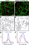
Planar polarity of ependymal basal body patches is disrupted by loss of primary cilia from radial glial progenitors. A, B, Confocal images of control and Nestin::Cre;Kif3afl/fl mutant lateral wall wholemounts stained for β-catenin (green) and γ-tubulin (red). Basal bodies in mutant ependymal cells were maintained in tightly clustered patches but the planar polarized position of these patches was less evident. Scale bar, 10 μm. C, D, Traces of the apical surfaces and basal body patches in A, B, with vectors indicating the relative position of the BB patch. Cells shaded in gray had BB patches that were not displaced from the cell center. E, Histogram showing the distribution of BB patch angles around the median in control (blue) and mutant (red) ependyma. Orientation of BB patch angles was dramatically disrupted in Nestin::Cre;Kif3afl/fl mutants compared with controls (curve fit test, p < 0.0001), also significantly more disrupted when compared directly with hGFAP::Cre;Kif3afl/fl mutants (p < 0.0001, see Fig. 3I red curve). F, Histogram showing the distribution of normalized BB patch displacement from center in control (blue) and mutant (red) ependyma (curve fit test, p < 0.0001). Nestin::Cre;Kif3afl/fl mutants had ependymal cells with more centrally positioned BB patches.
Figure 7.
Figure 7.
Summary illustration of the roles of primary and motile cilia in ependymal planar polarity. Normal development of ependymal planar polarity is shown in the left column. Loss of motile cilia from ependymal cells (middle column) does not dramatically alter translational polarity of basal body patches but it disrupts their rotational polarity, while loss of both primary cilia from radial glial progenitors and motile cilia from ependymal cells (right column) dramatically disrupts translational polarity.

Similar articles

Cited by

References

    1. Afzelius BA. Cilia-related diseases. J Pathol. 2004;204:470–477. - PMC - PubMed
    1. al-Shroof M, Karnik AM, Karnik AA, Longshore J, Sliman NA, Khan FA. Ciliary dyskinesia associated with hydrocephalus and mental retardation in a Jordanian family. Mayo Clin Proc. 2001;76:1219–1224. - PubMed
    1. Banizs B, Pike MM, Millican CL, Ferguson WB, Komlosi P, Sheetz J, Bell PD, Schwiebert EM, Yoder BK. Dysfunctional cilia lead to altered ependyma and choroid plexus function, and result in the formation of hydrocephalus. Development. 2005;132:5329–5339. - PubMed
    1. Boisvieux-Ulrich E, Laine MC, Sandoz D. The orientation of ciliary basal bodies in quail oviduct is related to the ciliary beating cycle commencement. Biol Cell. 1985;55:147–150. - PubMed
    1. Bruni JE. Ependymal development, proliferation, and functions: a review. Microsc Res Tech. 1998;41:2–13. - PubMed

Publication types

LinkOut - more resources