. 2010 Feb 17;30(7):2623-35.
doi: 10.1523/JNEUROSCI.5115-09.2010.

Suppression of the intrinsic apoptosis pathway by synaptic activity

Affiliations

Suppression of the intrinsic apoptosis pathway by synaptic activity

Frédéric Léveillé et al. J Neurosci. .

Abstract

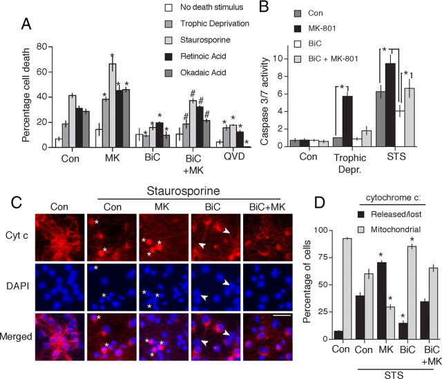
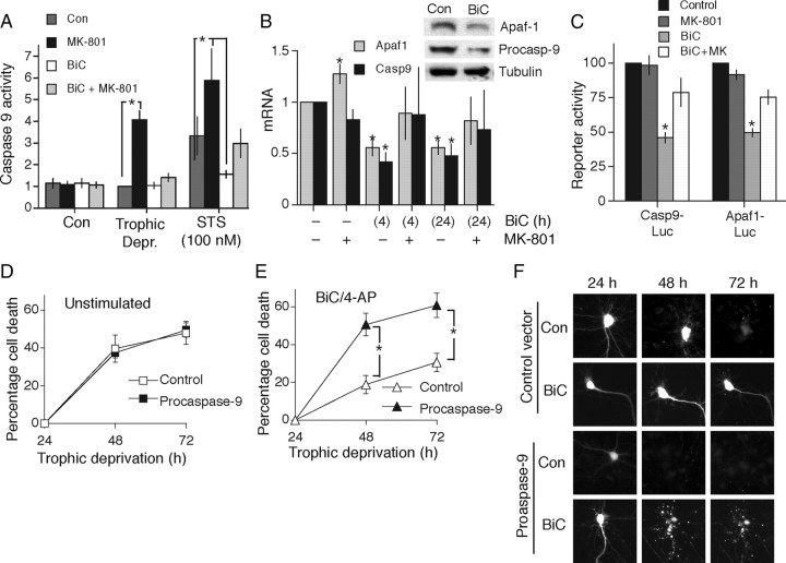
Synaptic activity promotes resistance to diverse apoptotic insults, the mechanism behind which is incompletely understood. We show here that a coordinated downregulation of core components of the intrinsic apoptosis pathway by neuronal activity forms a key part of the underlying mechanism. Activity-dependent protection against apoptotic insults is associated with inhibition of cytochrome c release in most but not all neurons, indicative of anti-apoptotic signaling both upstream and downstream of this step. We find that enhanced firing activity suppresses expression of the proapoptotic BH3-only member gene Puma in a NMDA receptor-dependent, p53-independent manner. Puma expression is sufficient to induce cytochrome c loss and neuronal apoptosis. Puma deficiency protects neurons against apoptosis and also occludes the protective effect of synaptic activity, while blockade of physiological NMDA receptor activity in the developing mouse brain induces neuronal apoptosis that is preceded by upregulation of Puma. However, enhanced activity can also confer resistance to Puma-induced apoptosis, acting downstream of cytochrome c release. This mechanism is mediated by transcriptional suppression of apoptosome components Apaf-1 and procaspase-9, and limiting caspase-9 activity, since overexpression of procaspase-9 accelerates the rate of apoptosis in active neurons back to control levels. Synaptic activity does not exert further significant anti-apoptotic effects downstream of caspase-9 activation, since an inducible form of caspase-9 overrides the protective effect of synaptic activity, despite activity-induced transcriptional suppression of caspase-3. Thus, suppression of apoptotic gene expression may synergize with other activity-dependent events such as enhancement of antioxidant defenses to promote neuronal survival.


Figures

Figure 1.
Figure 1.
Neuroprotection afforded by synaptic activity. A, Synaptic activity protects neurons against inducers of apoptosis. Mouse cortical neurons were treated in the presence or absence of the indicated compounds [BiC/4-AP: bicuculline (50 μm) plus 4-aminopyridine (250 μm) is labeled as “BiC” in figures]; MK-801 used at 10 μm here and throughout. These drugs were added 16 h before the addition of inducers of apoptosis (staurosporine, 100 nm; retinoic acid, 5 μm; okadaic acid, 2 nm). QVD-Oph was used at 50 μm and added 1 h before the addition of inducers of apoptosis. After a further 24 h, the neurons were fixed and levels of cell death calculated. In the case of trophic deprivation, neurons were subjected to 72 h in trophically deprived medium in the presence or absence of BiC, MK-801, or QVD-Oph. *p < 0.05 (compared to levels of death in the equivalent control condition); #p < 0.05 compared to levels of death in the BiC/4-AP-stimulated neurons [two-way ANOVA followed by Fisher's LSD test (n = 4)]. “No death stimulus” refers to neurons placed in standard transfection medium containing the insulin-transferrin-selenite supplement (trophic medium). B, Caspase-3/7 activation by apoptotic stimuli is suppressed by synaptic NMDAR activity. For the trophic deprivation model, neurons were treated with the indicated drugs for 48 h in trophically deprived medium. For the staurosporine (STS) model, neurons were treated with the indicated drugs for 16 h before treatment with staurosporine (100 nm) for a further 16 h. Control condition is trophic medium as described in A. Caspase-3/7 activity was measured and normalized to protein levels ascertained by BCA assay. *p < 0.05, one-way ANOVA followed by Fisher's LSD test here and elsewhere unless otherwise stated (n = 4). C, Synaptic NMDAR activity inhibits staurosporine-induced cytochrome c (Cyt c) release. Neurons were treated as in A. Cytochrome c immunofluorescent staining was performed 16 h after exposure to staurosporine. Asterisks highlight cells exhibiting diffuse staining throughout the neuron, a transient state that precedes loss of staining and apoptosis. The white arrows highlight neurons where cytochrome c has been lost, but nuclear chromatin fragmentation has not taken place, indicative of protection downstream of cytochrome c release. Pictures are representative of four independent experiments. Scale bar, 40 μm. D, Quantification of data shown in C. For each condition, ∼1500 cells were analyzed across four independent experiments. *p < 0.05 compared to control condition (STS treated).
Figure 2.
Figure 2.
Activity-dependent suppression of the proapoptotic gene Puma protects neurons. A, Activity-dependent suppression of the proapoptotic gene Puma. QRT-PCR analysis of Puma expression in cortical neurons treated with the indicated drugs. B, Western blot illustrating regulation of Puma expression at the protein level. C, The Puma promoter is sufficient to confer activity-dependent suppression on a reporter gene. Neurons were transfected with a Puma-luciferase reporter, plus a TK-Renilla control vector. At 24 h after transfection, neurons were stimulated for the indicated times, and firefly luciferase reporter activity was measured, normalized to the Renilla control. *p < 0.05, Student two-tailed t test (n = 4). D, E, Puma expression is sufficient to induce apoptosis in cortical neurons. Neurons were transfected with the indicated vectors, plus a vector encoding eGFP as a transfection marker. Transfected neurons were identified after 10 h, and their fate was monitored at 20 and 30 h. *p < 0.05 (n = 3).
Figure 3.
Figure 3.
Synaptic activity can suppress the proapoptotic effect of Puma upregulation. A, B, The neuroprotective effect of synaptic activity against staurosporine-induced apoptosis is occluded by Puma deficiency. Both wild-type and Puma-deficient neurons were stimulated as indicated for 16 h before the addition of staurosporine (100 nm) where indicated, for a further 24 h. After 24 h cell death was assessed. *p < 0.05 (n = 4) indicates stimulus-dependent differences (n = 4). #p < 0.05 indicates differences due to genotype (i.e., compared to +/+ neurons treated in the same way); “ns” indicates no significant statistical difference. B, Example phase-contrast pictures alongside the same field of cells after fixation and DAPI staining of nuclei. Scale bar, 50 μm. C, D, The neuroprotective effect of synaptic activity against trophic deprivation-induced apoptosis is occluded by Puma deficiency. Wild-type and Puma-deficient neurons were treated as indicated for 72 h in trophically deprived medium, after which cells were fixed and levels of cell death assessed. *p < 0.05 (n = 4). *p < 0.05 (n = 4) indicates stimulus-dependent differences (n = 4). #p < 0.05 indicates differences due to genotype (i.e., compared to +/+ neurons treated in the same way); “ns” indicates no significant statistical difference. D, Example phase-contrast pictures alongside the same field of cells after fixation and DAPI staining of nuclei. Scale bar, 50 μm. E, Synaptic activity can suppress the proapoptotic effect of Puma upregulation. Neurons were treated where indicated with BiC/4-AP starting 24 h before being transfected with peGFP plus vectors encoding either Puma, β-globin, or Puma-ΔBH3. Where used, QVD-Oph was applied to neurons 1 h before transfection. For each expression vector, and for each treatment, the number of viable neurons per field was analyzed at 10 h after transfection. The effect of BiC/4-AP-induced AP burst activity, or the effect of QVD-Oph, on the number of Puma-expressing neurons seen is expressed relative to the effect of these treatments on the number of neurons expressing the two control vectors (β globin and Puma-ΔBH3). *p < 0.05 (n = 6). F, G, Suppression of Puma-induced apoptosis by synaptic activity or caspase inhibition is downstream of cytochrome c release. Neurons are shown expressing eGFP plus either control vector (β-globin), Puma or Puma-ΔBH3. To inhibit Puma-induced apoptosis neurons were either pretreated with BiC/4-AP or QVD-Oph, as described in the text. At 10 h after transfection neurons were fixed and processed for cytochrome c immunofluorescence. G, Quantification of the percentage of viable Puma and Puma ΔBH3-expressing neurons exhibiting cytochrome c release at 10 h after transfection. 60–100 cells were analyzed for each treatment across three independent experiments.
Figure 4.
Figure 4.
Synaptic activity suppresses the expression of Apaf-1 and procaspase-9. A, Synaptic NMDAR activity inhibits activation of caspase-9 in response to an apoptotic insult. Neurons were treated with the indicated compounds as described in Figure 1A. Caspase-9 activity was measured after 48 h trophic deprivation, or 16 h staurosporine treatment. *p < 0.05 (n = 4). B, Synaptic NMDAR activity suppresses expression of apoptosome components. QRT-PCR analysis of Apaf1 and Casp9 expression in neurons. *p < 0.05 (n = 4). Inset shows example Western blot illustrating BiC/4-AP-induced suppression of protein expression. C, Promoter regions of Apaf1 and Casp9 confer activity-dependent suppression on a luciferase reporter gene. Neurons were transfected with the indicated reporters, plus a TK-Renilla control vector. At 24 h after transfection, neurons were stimulated for the indicated times and firefly luciferase reporter activity was measured, normalized to the Renilla control. *p < 0.05 (n = 3). D, E, Overexpression of procaspase-9 accelerates apoptosis in AP-bursting but not control neurons. Neurons expressing eGFP plus either a control vector (β globin) or a vector encoding procaspase-9 were placed in trophically deprived medium and either left unstimulated (D) or treated with BiC/4-AP to induce AP bursting (E). Pictures of neurons were taken at 24 h and then their viability was monitored at 24 h intervals. *p < 0.05 (n = 5). F, Example pictures relating to D and E.
Figure 5.
Figure 5.
Synaptic activity does not exert protection downstream of caspase-9 activation. A, QRT-PCR analysis of Casp3 expression in neurons stimulated as indicated. *p < 0.05 (n = 3). B, The inducible caspase-9 system enables neurons to be killed by treatment with the dimerizer drug AP20187. Neurons were transfected with peGFP plus either control or icaspase-9 encoding vectors. At 24 h after transfection pictures of neurons were taken before the treatment, where indicated, with AP20187 (100 pm), to cause dimerization/activation of icaspase-9. Cell death was assessed after a further 24 h. Where used, QVD-Oph was applied 1 h before AP20187. *p < 0.05 (n = 4). Inset shows example pictures. C, Synaptic activity cannot protect against icaspase-9-induced apoptosis. Neurons were transfected as in B and immediately left either unstimulated or treated with BiC/4-AP to induce burst activity. At 24 h after transfection pictures of cells were taken before treatment with the indicated concentrations of AP20187. Cell death was assessed after a further 24 h (n = 4).
Figure 6.
Figure 6.
Neuroprotection due to activity-dependent suppression of Puma, Apaf-1, and Procaspase-9 expression is not via p53 inhibition. A, QRT-PCR analysis of p53 expression in p53 +/+ and +/− neurons that were stimulated as indicated for 4 h. *p < 0.05 (n = 6 +/+, n = 4 +/−). B, QRT-PCR analysis of Puma expression in p53 +/+, +/−, and −/− neurons that were stimulated as indicated. MK-801 treatment was for 24 h. BiC/4-AP treatment was for 4 h. *p < 0.05 (n = 6 +/+, n = 4 +/−, n = 4 −/−) indicates stimulus-dependent differences (n = 4). #p < 0.05 indicates differences due to genotype (i.e., compared to +/+ neurons treated in the same way). @p < 0.05 compared to level of neuronal death in BiC/4-AP-stimulated, p53 +/+ neurons. C, QRT-PCR analysis of Apaf1 and Casp9 expression in p53 +/+, +/−, and −/− neurons stimulated where indicated with BiC/4-AP for 4 h. #p < 0.05 compared to control of that particular genotype (n = 6 +/+, n = 4 +/−, n = 4 −/−). D, The neuroprotective effect of synaptic activity against staurosporine-induced apoptosis is not occluded by p53 deficiency. p53 +/+, +/−, and −/− neurons were stimulated as indicated for 16 h before the addition of staurosporine (100 nm) where indicated, for a further 24 h. After 24 h, cell death was assessed. *p < 0.05 (n = 6 +/+, n = 4 +/−, n = 4 −/−) indicates stimulus-dependent differences (n = 4). #p < 0.05 indicates differences due to genotype (i.e., compared to +/+ neurons treated in the same way). E, The neuroprotective effect of synaptic activity against trophic deprivation-induced apoptosis is not occluded by p53 deficiency. p53 +/+, +/−, and −/− neurons were stimulated as indicated for 72 h in trophically deprived medium, after which cell death was assessed. *p < 0.05 (n = 6 +/+, n = 4 +/−, n = 4 −/−) indicates stimulus-dependent differences (n = 4). #p < 0.05 indicates differences due to genotype (i.e., compared to +/+ neurons treated in the same way). There are no such differences.

Similar articles

Cited by

References

    1. Adams SM, de Rivero Vaccari JC, Corriveau RA. Pronounced cell death in the absence of NMDA receptors in the developing somatosensory thalamus. J Neurosci. 2004;24:9441–9450. - PMC - PubMed
    1. Akhtar RS, Geng Y, Klocke BJ, Latham CB, Villunger A, Michalak EM, Strasser A, Carroll SL, Roth KA. BH3-only proapoptotic Bcl-2 family members Noxa and Puma mediate neural precursor cell death. J Neurosci. 2006;26:7257–7264. - PMC - PubMed
    1. Al-Mubarak B, Soriano FX, Hardingham GE. Synaptic NMDAR activity suppresses FOXO1 expression via a cis-acting FOXO binding site: FOXO1 is a FOXO target gene. Channels (Austin) 2009;3:233–238. - PMC - PubMed
    1. Bading H, Ginty DD, Greenberg ME. Regulation of gene expression in hippocampal neurons by distinct calcium signaling pathways. Science. 1993;260:181–186. - PubMed
    1. Benn SC, Woolf CJ. Adult neuron survival strategies—slamming on the brakes. Nat Rev Neurosci. 2004;5:686–700. - PubMed

Publication types

MeSH terms