. 2010 Feb 17;30(7):2636-49.
doi: 10.1523/JNEUROSCI.4456-09.2010.

Amyloid beta induces the morphological neurodegenerative triad of spine loss, dendritic simplification, and neuritic dystrophies through calcineurin activation

Affiliations

Amyloid beta induces the morphological neurodegenerative triad of spine loss, dendritic simplification, and neuritic dystrophies through calcineurin activation

Hai-Yan Wu et al. J Neurosci. .

Abstract

Amyloid beta (Abeta)-containing plaques are surrounded by dystrophic neurites in the Alzheimer's disease (AD) brain, but whether and how plaques induce these neuritic abnormalities remain unknown. We tested the hypothesis that soluble oligomeric assemblies of Abeta, which surround plaques, induce calcium-mediated secondary cascades that lead to dystrophic changes in local neurites. We show that soluble Abeta oligomers lead to activation of the calcium-dependent phosphatase calcineurin (CaN) (PP2B), which in turn activates the transcriptional factor nuclear factor of activated T cells (NFAT). Activation of these signaling pathways, even in the absence of Abeta, is sufficient to produce a virtual phenocopy of Abeta-induced dystrophic neurites, dendritic simplification, and dendritic spine loss in both neurons in culture and in the adult mouse brain. Importantly, the morphological deficits in the vicinity of Abeta deposits in a mouse model of AD are ameliorated by CaN inhibition, supporting the hypothesis that CaN-NFAT are aberrantly activated by Abeta and that CaN-NFAT activation is responsible for disruption of neuronal structure near plaques. In accord with this, we also detect increased levels of an active form of CaN and NFATc4 in the nuclear fraction from the cortex of patients with AD. Thus, Abeta appears to mediate the neurodegeneration of AD, at least in part, by activation of CaN and subsequent NFAT-mediated downstream cascades.


Figures
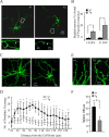
Figure 1.
Figure 1.
Abnormal morphologies in neurons from Tg cultures. A, Representative images of a wild-type GFP-labeled neuron at 14 DIV shows intricately branched dendritic arbors (A1, A3), whereas Tg neurons exhibit simplified dendritic complexity and localized dendritic dystrophies (A2, A4). B, The percentage of neurons with dendritic dystrophies at 14 and 21 DIV was increased in Tg neurons compared with wild type (14 DIV: wild-type cultures, 2.0 ± 0.2%; Tg cultures, 14.0 ± 2.0%, p < 0.0001; 21 DIV: wild-type cultures, 3.0 ± 0.2%; Tg cultures, 24.0 ± 5.0%; p < 0.0001, wild-type vs Tg; p = 0.0003, 14 vs 21 DIV). n = 50 cells from each condition. C, Representative images of wild-type or Tg GFP-labeled mature neurons (21 DIV). D, Sholl analysis of total number of branch point on basal dendrites of neuron from wild-type and Tg cultures shows decreased complexity in Tg neurons starting at 30 μm from the cell body. n = 25 cells from each condition. E, Representative GFP-labeled dendritic segments studded with mature spines from wild-type and Tg neurons show a loss of spines on Tg dendrites that is confirmed in quantitative assay of spine densities determined in neurons without apparent dystrophies at 21 DIV in wild-type and Tg cultures (F). n = 4 culture per experiment and 400 spines from each condition. *p < 0.05; **p < 0.01; data represent mean ± SD.
Figure 2.
Figure 2.
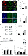
Aβ induces NFATc4 and CaN-aberrant nuclear localization in Tg neurons in culture and AD postmortem brains. A, Immunostaining images of NFATc4 in Tg or wild-type neurons. Neurons from Tg or wild-type culture at 14 DIV are labeled with NFATc4 (red), MAP2 (green), and Hoechst nuclear counterstain (blue). Compared with wild-type neurons, Tg neurons show higher immunoreactivity of NFATc4 in the nuclei. B, Quantification of NFATc4 nuclear immunoreactivity obtained from A. n = 40 cells from 3 different experiments. * p < 0.05; data represent mean ± SD. C, Immunostaining images of CaN in Tg or wild-type neurons. Neurons from Tg or wild-type culture at 14 DIV are labeled with CaN (green) and Hoechst nuclear counterstain (blue). Compared with wild-type neurons, Tg neurons show higher immunoreactivity of CaN in the nuclei. D, Quantification of CaN nuclear immunoreactivity obtained from C. n = 40. *p < 0.01; data represent mean ± SD. E, F, Subcellular fractions were prepared from brains of AD patients or controls and analyzed by immunoblotting for NFATc4 and cytoplasmic or nuclear control proteins GAPDH and HDAC1, respectively. G, Semiquantification of NFATc4 immunoreactivity in E. NFATc4 was detected in all three compartments but substantially enriched in nuclei in AD brains (n = 6 for each condition). *p < 0.05; data represent mean ± SD. H, I, Semiquantification of immunoreactivity from each subcellular fraction for both full-length CaN (60 kDa) (H) and CaNCA (45 kDa) (I). Inset in I shows representative blots of CaN in nuclear fraction. *p < 0.05; data represent mean ± SEM.
Figure 3.
Figure 3.
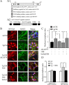
Conditioned medium induces NFATc4-aberrant nuclear translocation in wild-type cultured neurons. A, Schematic representation of AKAP79 inhibitory peptide for CaN (A1) and AAV2 viral vectors (AAV–CMV/CBA–WPRE) with AKAP79 inhibitory peptide (A2). B, Aβ depletion or CaN inhibition with AKAP79 inhibitory peptide prevents TgCM-induced NFATc4 activation as seen in cultures stained with NFATc4 (red), MAP2 (green), and Hoechst nuclear counterstain (blue) from each experimental condition. C, Quantification of NFATc4-aberrant nuclear translocation induced by TgCM in wild-type cultured neurons. The ratio of nucleus to cytoplasm is shown. TgCM causes an increase in the nucleus/cytoplasm ratio of NFATc4 that is dependent on the presence of Aβ (because anti-Aβ antibody 3D6 prevents the effect). Inhibition of CaN activation by AKAP79 peptide also prevented the increase in the nucleus/cytoplasm ratio of NFATc4 (n > 40 cells). Data represent mean ± SD. *p < 0.05. D, Quantification of NFATc4-aberrant nuclear translocation induced by different SEC fractions in wild-type cultured neurons. Application of SEC fractions 6–7 (sAPP fraction) from either TgCM or wtCM caused no significant difference on the nucleus/cytoplasm ratio of NFATc4. However, application of SEC fractions 18–19 (Aβ fraction) of TgCM onto wild-type neurons for 24 h caused significant increase in translocation of NFATc4 to the nucleus, but no changes were observed in neurons applied with the same SEC fractions of wtCM. Immunodepletion of Aβ from the fractions 18–19 of TgCM with 3D6 prevented the increase in the nucleus/cytoplasm ratio of NFATc4. n > 35 cells. Data represent mean ± SD; *p < 0.05. ITR, Inverted terminal repeat.
Figure 4.
Figure 4.
Aberrant neuronal morphologies induced by TgCM or APP overexpression are prevented by Aβ depletion. A, Primary cultures were maintained in TgCM for 21 DIV in different culture conditions, as indicated. The percentage of neurons with dendritic dystrophies at 21 DIV is increased in the presence of TgCM, and this beading can be prevented by immunodepletion of Aβ. B, C, Representative GFP-labeled mature neurons (B) and Sholl analysis of branching on dendrites of neurons from indicated conditions shows that TgCM reduced branching and that this is rescued by immunodepletion of Aβ with 3D6 (C). D, Spine densities are determined in neurons without apparent dystrophies in each experimental condition, showing a decrease in spine density with TgCM that could be rescued with 3D6 treatment. E–H, Aβ depletion prevents APP overexpression-induced morphological abnormalities. In Tg cultures at 21 DIV, dendritic dystrophies (E), dendritic attenuation (F, G) and spine loss (H) are prevented by Aβ depletion with 3D6. Sixty cells were analyzed per experimental condition in each experiment. *p < 0.05; **p < 0.01; *p < 0.05 in G (Tg with 3D6 vs Tg with boiled 3D6). Data represent mean ± SD.
Figure 5.
Figure 5.
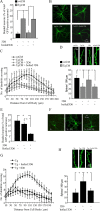
CaN inhibition by AKAP79 inhibitory peptide prevents TgCM-induced morphological abnormalities. Dendritic dystrophies (A), dendritic attenuation (B, C), and spine loss (D) in wild-type cultured neurons growing in TgCM are significantly prevented by CaN inhibitory peptide AKAP79. *p < 0.05 in C (TgCM with AKAP79 vs TgCM with vector). **p < 0.01. Data are mean ± SD from three independent experiments in triplicate. E–H, CaN inhibition by AKAP79 inhibitory peptide prevents APP overexpression-induced morphological abnormalities. In Tg cultures at 21 DIV, dendritic dystrophies (E), dendritic attenuation (F, G), and spine loss (H) are prevented by CaN inhibitory peptide AKAP79. *p < 0.05 in G (Tg with AKAP79 vs Tg with vector). Data are mean ± SD from three independent experiments, each in triplicate.
Figure 6.
Figure 6.
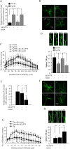
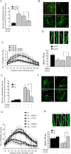
Inhibition of CaN–NFAT interaction by VIVIT prevents TgCM-induced morphological abnormalities. Dendritic dystrophies (A), dendritic attenuation (B, C), and spine loss (D) in wild-type cultured neurons growing in TgCM are significantly prevented by VIVIT (catalog #480401; Calbiochem). *p < 0.05 in C (TgCM with VIVIT vs TgCM with DMSO). Data are mean ± SD from three independent experiments in triplicate. E–H, Inhibition of CaN–NFAT interaction by VIVIT prevents APP overexpression-induced morphological abnormalities. In Tg cultures at 21 DIV, dendritic dystrophies (E), dendritic attenuation (F, G), and spine loss (H) are prevented by VIVIT. *p < 0.05 in G (Tg with VIVIT vs Tg with DMSO). Data are mean ± SD from three independent experiments in triplicate.
Figure 7.
Figure 7.
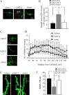
Wild-type cultured neurons overexpressing a constitutively active CaN construct, CaNCA, develop abnormal morphology that is a phenocopy of the Aβ effect. Overexpression of CaNCA, but not CaNwt or vector control, into wild-type cultures induces dendritic dystrophies (A, B; arrows indicated local swelling of dendrites), simplification of dendritic arborization (C, D), and spine loss (E, F). Neurons expressing CaNCA displayed significantly more neurons with dendritic dystrophies, simplified dendritic complexity, and spine loss than either CaNwt or vector-expressing neurons. *p < 0.05; *p < 0.05 in D (CaNwt vs CaNCA). Data are mean ± SD from three independent experiments, each in triplicate.
Figure 8.
Figure 8.
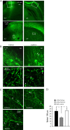
Overexpression of CaNCA, but not CaNwt, in the intact mouse brain induces abnormal morphologies. A, Representative images in different magnifications from cortex (A1, A3) and hippocampal areas (A2, A4) after CaNCA or CaNwt transduction showing GFP expression in fixed postmortem brain sections stained with GFP antibody. B, Representative high-power images of neuronal processes corresponding to areas indicated in A from CaNwt (B1, B3) or CaNCA (B2, B4) injected mice. Arrows indicated neurites or axons with typical dystrophies. C, D, Images of dendritic spine (C) and quantitative analysis of spine densities (D) from live imaging of neurons without apparent dystrophies shows a decrease in spine density with CaNCA overexpression. In C, arrows indicated spines, and arrowhead indicated neurites with typical dystrophies. *p < 0.05; values represent mean ± SD (n = 4 animals for each condition and total >400 spines).
Figure 9.
Figure 9.
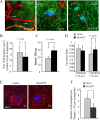
Abnormal morphologies are prevented by overexpression of CaN inhibitory peptide AKAP79 in APP/PS1 mouse brain. A1, In vivo low-magnification image of GFP-expressing neurites and axons (green), blood vessels containing Texas Red, and amyloid deposition stained with methoxy-XO4 (blue). Arrows indicated Aβ deposits. A2, A3, High-magnification live images taken from ∼100 μm below the brain surface in the neocortex of adult APP/PS1 mice show dendritic spines and dystrophies in both vector- and AKAP79-expressing conditions. B–D, Quantification of spine density (B), dendritic dystrophies (C), and neurite curvature (D) near Aβ deposits in vector- or AKAP79-expressing APP/PS1 mouse brain shows that AKAP79 expression decreases dystrophy size (B; vector, 16.4 ± 6.1 μm2; AKAP79, 12.8 ± 3.6; p = 0.029; n = 45 from 4 animals), increases spine density (C; vector, 17.8 ± 3.0/100 μm; AKAP79, 28.3 ± 6.1; p < 0.001; n > 400 spines), and decreases abnormal neurite curvature near plaques (D) (n = 4 animals for each condition and total > 400 spines). E, Postmortem sections indicated axonal dystrophies with SMI312 staining (red) and plaques with thioflavin S (blue) shows that plaque-associated axonal dystrophies are reduced (quantified in F; vector, 16.4 ± 6.1 μm2; AKAP79, 12.8 ± 3.6; p = 0.0291; n = 45 plaques from 4 animals) in areas injected with AKAP79. F, Quantification of numbers of axonal dystrophies per single Aβ deposits from postmortem sections. Values represent mean ± SD. *p < 0.05; **p < 0.01.

Similar articles

Cited by

References

    1. Abdul HM, Sama MA, Furman JL, Mathis DM, Beckett TL, Weidner AM, Patel ES, Baig I, Murphy MP, LeVine H, Jr, Kraner SD, Norris CM. Cognitive decline in Alzheimer's disease is associated with selective changes in calcineurin/NFAT signaling. J Neurosci. 2009;29:12957–12969. - PMC - PubMed
    1. Agostinho P, Lopes JP, Velez Z, Oliveira CR. Overactivation of calcineurin induced by amyloid-beta and prion proteins. Neurochem Int. 2008;52:1226–1233. - PubMed
    1. Aramburu J, Yaffe MB, López-Rodríguez C, Cantley LC, Hogan PG, Rao A. Affinity-driven peptide selection of an NFAT inhibitor more selective than cyclosporin A. Science. 1999;285:2129–2133. - PubMed
    1. Bacskai BJ, Hickey GA, Skoch J, Kajdasz ST, Wang Y, Huang GF, Mathis CA, Klunk WE, Hyman BT. Four-dimensional multiphoton imaging of brain entry, amyloid binding, and clearance of an amyloid-beta ligand in transgenic mice. Proc Natl Acad Sci U S A. 2003;100:12462–12467. - PMC - PubMed
    1. Berridge MJ, Lipp P, Bootman MD. The versatility and universality of calcium signalling. Nat Rev Mol Cell Biol. 2000;1:11–21. - PubMed

Publication types

MeSH terms

LinkOut - more resources