. 2010 Feb 17;30(7):2700-9.
doi: 10.1523/JNEUROSCI.5011-09.2010.

Saccade suppression by electrical microstimulation in monkey caudate nucleus

Affiliations

Saccade suppression by electrical microstimulation in monkey caudate nucleus

Masayuki Watanabe et al. J Neurosci. .

Abstract

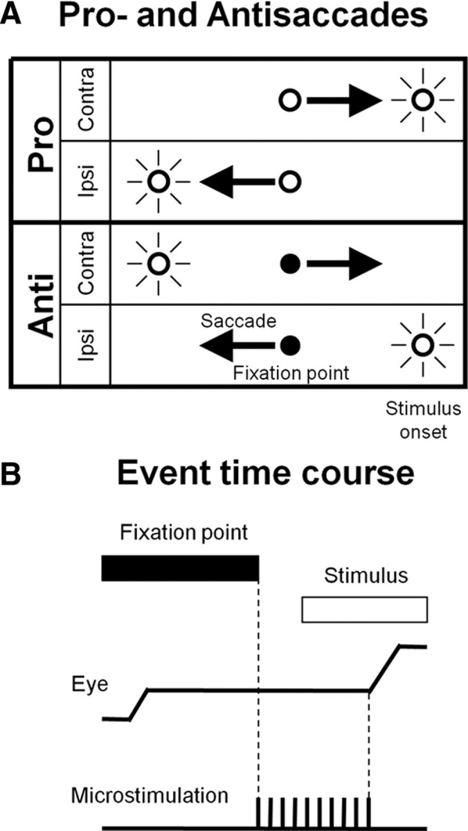
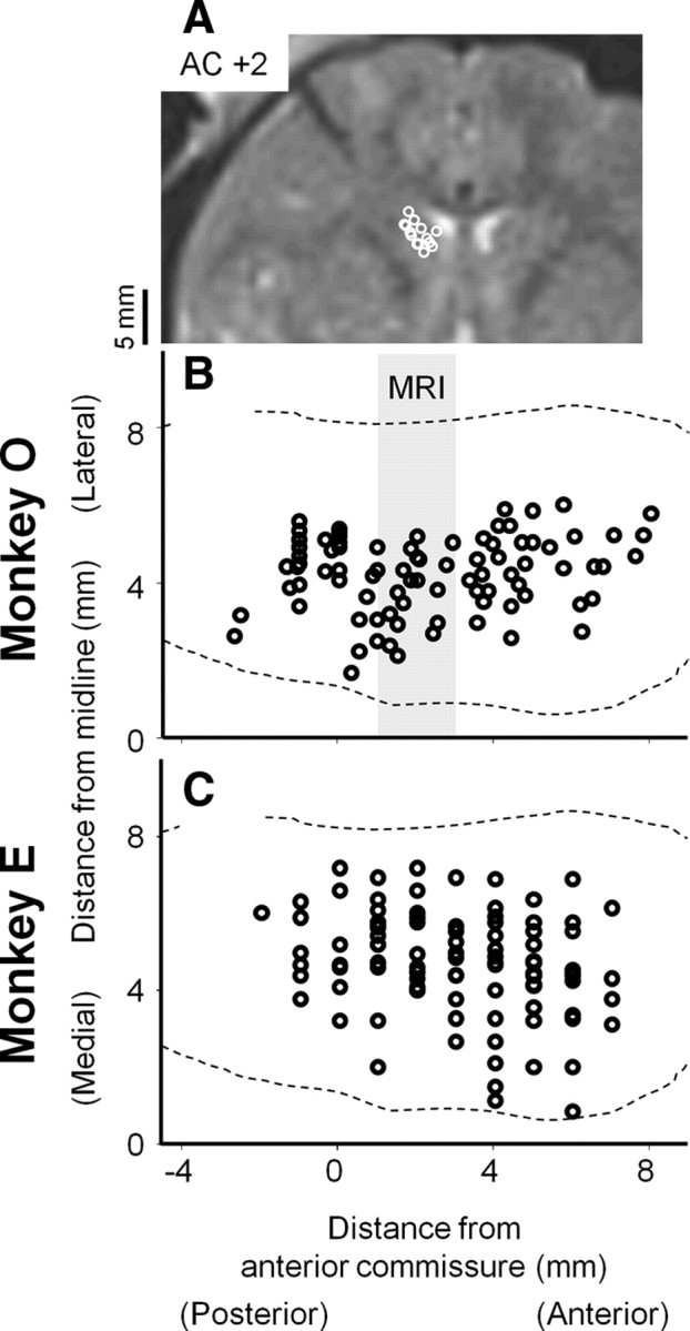
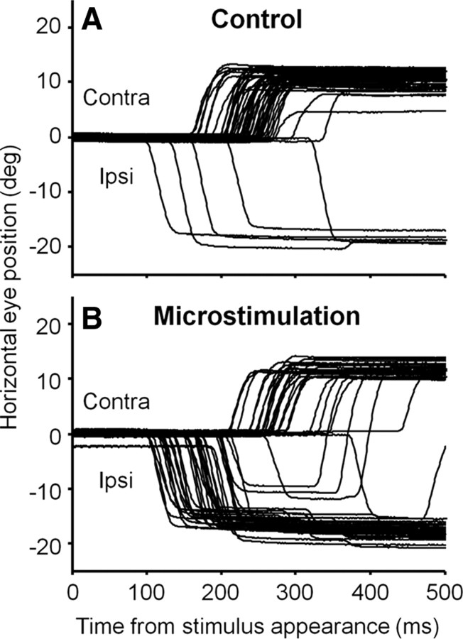
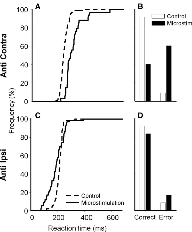
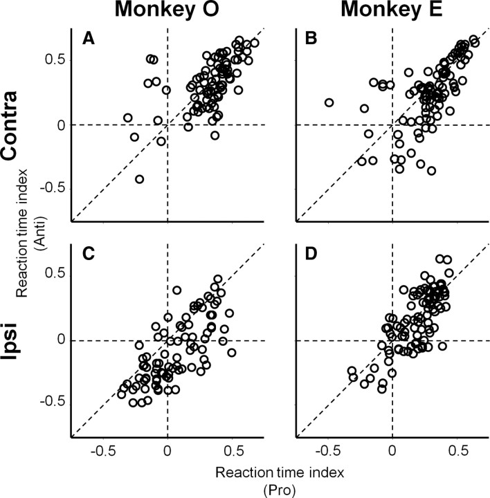
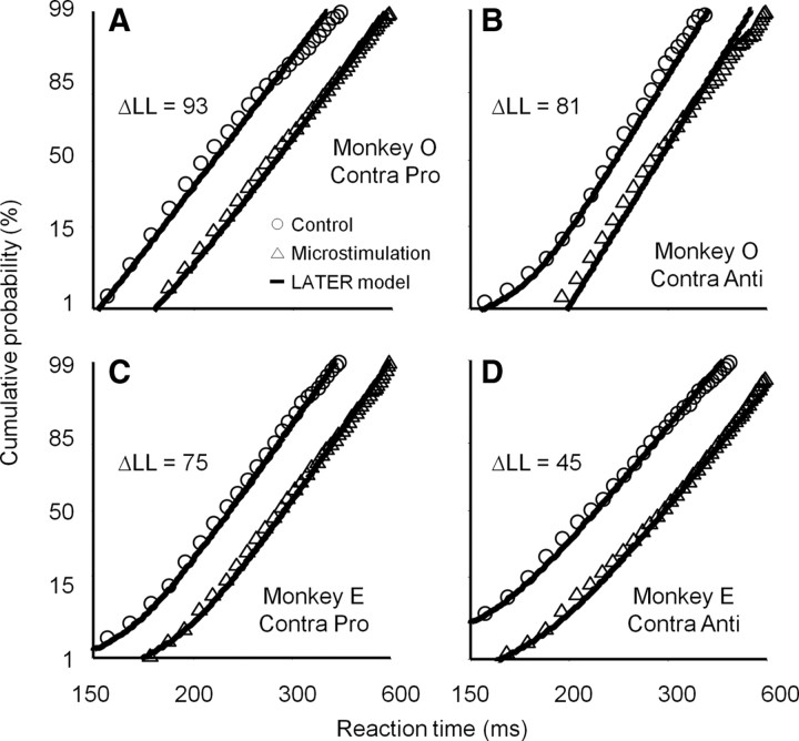
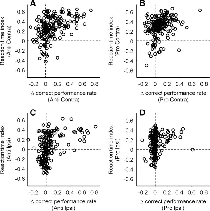
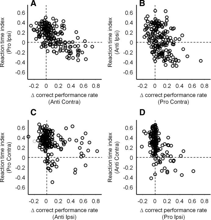
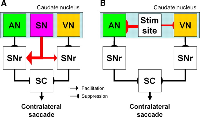
It has been suggested that the caudate nucleus, the input stage of the basal ganglia, facilitates and suppresses saccade initiation based on its anatomical characteristics. Although the involvement of the caudate nucleus in saccade facilitation has been shown previously, it is still unclear whether the caudate nucleus is also involved in saccade suppression. Here, we revealed the direct involvement of the caudate nucleus in saccade suppression by electrical microstimulation in behaving monkeys. We delivered microstimulation to the caudate nucleus while monkeys performed the prosaccade (look toward a peripheral visual stimulus) and antisaccade (look away from the stimulus) paradigm. The reaction times of contralateral saccades were prolonged on both prosaccade and antisaccade trials. The suppression effects on reaction times were stronger on prosaccade trials compared with antisaccade trials. The analysis of reaction time distributions using the linear approach to threshold with ergodic rate model (LATER model) revealed that microstimulation prolonged reaction times by reducing the rate of rise to the threshold for saccade initiation. Microstimulation also worsened correct performance rates for contralateral saccades. The same microstimulation prolonged and/or shortened the reaction times of ipsilateral saccades, although the effects were not as consistent as those on contralateral saccades. We conclude that caudate signals are sufficient to suppress contralateral saccades and influence saccadic decision by controlling contralateral and ipsilateral saccade commands at the same time.


Figures

Figure 1.
Figure 1.
Prosaccade and antisaccade paradigm. A, Four task conditions. Fixation point color indicates monkeys to perform a prosaccade (look toward a stimulus) or an antisaccade (look away from the stimulus). “Contra” and “Ipsi” indicate saccade directions. B, Event time course. After fixation point appearance, monkeys acquire the fixation point and generate a saccade in response to stimulus appearance. The fixation point disappeared 200 ms before stimulus appearance. On randomly chosen 50% of trials, microstimulation was delivered from 200 ms before stimulus appearance to saccade initiation.
Figure 2.
Figure 2.
Reconstructed stimulation sites. A, MRI image at 2 mm anterior from the anterior commissure in monkey O. B, C, reconstructed site projected on the horizontal plane in monkey O (n = 85) (B) and E (n = 94) (C), respectively. Sites included in the gray stripes labeled as MRI in B are superimposed on the MRI image (A). Broken lines indicate the boundaries of the caudate nucleus (François et al., 1996). In monkey E, the level of the anterior commissure is estimated at 19 mm anterior from the intermeatal line (Mikula et al., 2007).
Figure 3.
Figure 3.
Effects of microstimulation on contralateral antisaccades at an example stimulation site in monkey O. A, Control trials. B, Microstimulation trials. Each trace indicates the time course of horizontal eye positions. Upward and downward deflections indicate correct (contralateral) and direction error (ipsilateral) saccades, respectively. The eye position traces are aligned with stimulus appearance.
Figure 4.
Figure 4.
Quantitative analyses of microstimulation effects on antisaccades at the same stimulation site in Figure 3. A, B, Cumulative reaction time distributions (A) and correct and direction error rates (B) on contralateral antisaccade trials. C, D, Ipsilateral antisaccade trials. Dotted and continuous lines in A and C indicate control and microstimulation trials, respectively. White and black bars in B and D indicate control and microstimulation trials, respectively. “Error” in B and D indicates direction error trials. No saccade trials are not included in this figure because they were not observed during this experiment.
Figure 5.
Figure 5.
Summary of microstimulation effects on saccade reaction times. A, Contralateral saccades in monkey O (n = 85). B, Contralateral saccades in monkey E (n = 94). C, Ipsilateral saccades in monkey O (n = 85). D, Ipsilateral saccades in monkey E (n = 94). x- and y-axes indicate reaction time indices for prosaccades and antisaccades, respectively. Positive and negative values of reaction time indices indicate prolonged and shortened reaction times by microstimulation, respectively.
Figure 6.
Figure 6.
LATER model for contralateral saccades with alteration in rate of rise. A, Prosaccade trials in monkey O. B, Antisaccade trials in monkey O. C, Prosaccade trials in monkey E. D, Antisaccade trials in monkey E. Stimulation sites in which microstimulation prolonged reaction times (t test p < 0.05) were collapsed. Circles and triangles indicate cumulative distributions of reaction times with 10 ms bin width on control and microstimulation trials, respectively. Continuous lines indicate the results of LATER model fittings under the constraint of alteration in the rate of rise to the threshold for saccade initiation. Fitting results under the constraint of alteration in the distance between the baseline and threshold are shown in supplemental Figure 3, available at www.jneurosci.org as supplemental material. The summary of fitting results is shown in Table 1. See also supplemental Figures 4 and 5, available at www.jneurosci.org as supplemental material, for the fitting results of ipsilateral saccades.
Figure 7.
Figure 7.
Summary of microstimulation effects on correct performance rates. A, Correlation between Δ correct performance rates and reaction time indices on contralateral antisaccade trials. B, Contralateral prosaccade trials. C, Ipsilateral antisaccade trials. D, Ipsilateral prosaccade trials. The number of data points in all panels is 179 (85 and 94 in monkeys O and E, respectively). Positive and negative values of Δ correct performance rates indicate worsened and improved performance by microstimulation, respectively. Positive correlation between Δ correct performance rates and reaction time indices indicates that correct performance rates were worsened when microstimulation prolonged saccade reaction times. We found the same positive correlations using Spearman's (nonparametric) correlation coefficients (data not shown). We confirmed similar results when data from two monkeys were analyzed separately.
Figure 8.
Figure 8.
Relationships between microstimulation effects on correct performance rates and reaction times for saccades toward the opposite directions in response to the same stimulus. A, Correlation between Δ correct performance rates on contralateral antisaccade trials and reaction times indices on ipsilateral prosaccade trials. B, Correct performance rates for contralateral prosaccades and reaction time indices for ipsilateral antisaccades. C, Correct performance rates for ipsilateral antisaccades and reaction time indices for contralateral prosaccades. D, Correct performance rates for ipsilateral prosaccades and reaction time indices for contralateral antisaccades. The number of data points in all panels is 179 (85 and 94 in monkeys O and E, respectively). Negative correlation between Δ correct performance rates and reaction time indices indicates that correct performance rates were worsened when microstimulation facilitated saccades toward the opposite direction. We confirmed the results using Spearman's correlation coefficients (data not shown). We also confirmed similar results when data from two monkeys were analyzed separately.
Figure 9.
Figure 9.
Hypothetical schematic diagram for contralateral saccades. There are three types of caudate neurons encoding different saccade signals. ANs encode predominantly automatic saccade commands toward the contralateral stimulus. VNs encode predominantly voluntary saccade commands toward the contralateral direction regardless of stimulus locations. SNs encode similar saccade commands with VNs, except that they show strong activation on ipsilateral antisaccade trials. ANs and VNs give rise to the facilitation (direct) pathway and suppress the tonic activity of SNr neurons to release the SC for contralateral saccade initiation. In contrast, SNs give rise to the suppression (indirect) pathway and activate SNr neurons to suppress contralateral saccade initiation. To account for the suppression effects of microstimulation on contralateral saccades, we speculate that (A) signals carried by the suppression pathway originated from SNs (indicated by red arrows) are stronger than those carried by the facilitation pathways originated from ANs and VNs. B, Another possibility is that microstimulation activated lateral inhibitory interactions within the caudate nucleus and suppressed the activity of ANs and VNs remote from the stimulation site. To account for the stronger suppression effects of microstimulation on prosaccade reaction times compared with antisaccade reaction times, we hypothesize that the suppression effects on ANs are stronger than those on VNs in both A and B, described by the thickness of the arrows. The suppression pathways are shown by single arrows for simplicity even though they are polysynaptic. Pathways from the SNr to cortex are omitted for simplicity.

Similar articles

Cited by

References

    1. Bahill AT, Clark MR, Stark L. The main sequence, a tool for studying human eye movements. Math Biosci. 1975;24:191–204.
    1. Bell AH, Everling S, Munoz DP. Influence of stimulus eccentricity and direction on characteristics of pro- and antisaccades in non-human primates. J Neurophysiol. 2000;84:2595–2604. - PubMed
    1. Briand KA, Strallow D, Hening W, Poizner H, Sereno AB. Control of voluntary and reflexive saccades in parkinson's disease. Exp Brain Res. 1999;129:38–48. - PubMed
    1. Carpenter RH, Williams ML. Neural computation of log likelihood in control of saccadic eye movements. Nature. 1995;377:59–62. - PubMed
    1. Cebrián C, Parent A, Prensa L. Patterns of axonal branching of neurons of the substantia nigra pars reticulata and pars lateralis in the rat. J Comp Neurol. 2005;492:349–369. - PubMed

Publication types

MeSH terms

LinkOut - more resources