. 2010 May;298(5):C1151-8.
doi: 10.1152/ajpcell.00382.2009. Epub 2010 Feb 17.

The contribution of AKAP5 in amylase secretion from mouse parotid acini

Affiliations

The contribution of AKAP5 in amylase secretion from mouse parotid acini

Ching-Yi Wu et al. Am J Physiol Cell Physiol. 2010 May.

Abstract

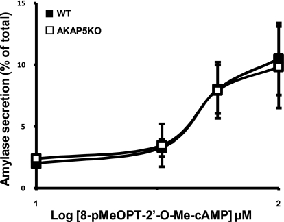
A-kinase (PKA) anchoring proteins (AKAPs) are essential for targeting type II PKA to specific locales in the cell to control function. In the present study, AKAP5 (formerly AKAP150) and AKAP6 were identified in mouse parotid acini by type II PKA regulatory subunit (RII) overlay assay and Western blot analysis of mouse parotid cellular fractions, and the role of AKAP5 in mouse parotid acinar cell secretion was determined. Mice were euthanized with CO(2). Immunofluorescence staining of acinar cells localized AKAP5 to the basolateral membrane, whereas AKAP6 was associated with the perinuclear region. In functional studies, amylase secretion from acinar cells of AKAP5 mutant [knockout (KO)] mice treated with the beta-adrenergic agonist, isoproterenol, was reduced overall by 30-40% compared with wild-type (WT) mice. In contrast, amylase secretion in response to the adenylyl cyclase (AC) activator, forskolin, and the cAMP-dependent protein kinase (PKA) activator, N(6)-phenyl-cAMP, was not statistically different in acini from WT and AKAP5 KO mice. Treatment of acini with isoproterenol mimicked the effect of the Epac activator, 8-(4-methoxyphenylthio)-2'-O-methyladenosine-3',5'-cyclic monophosphate (8-pMeOPT-2'-O-Me-cAMP), in stimulating Rap1. However, in contrast to isoproterenol, treatment of acini with 8-pMeOPT-2'-O-Me-cAMP resulted in stimulation of amylase secretion from both AKAP5 KO and WT acinar cells. As a scaffolding protein, AKAP5 was found to coimmunoprecipitate with AC6, but not AC8. Data suggest that isoproterenol-stimulated amylase secretion occurs via both an AKAP5/AC6/PKA complex and a PKA-independent, Epac pathway in mouse parotid acini.


Figures

Fig. 1.
Fig. 1.
A: identification of A-kinase anchoring proteins (AKAPs) in mouse parotid acini by type II PKA regulatory subunit (RII) overlay assay of cellular fractions enriched in cytoskeleton (Ck), membrane skeleton (Msk), soluble fraction (Sn), and cAMP pull-down isolates (PD), in the absence and presence of St-Ht31 (50 μM). Intracellular proteins that are likely to be AKAPs are indicated by arrows. Among these proteins, a and b were identified as AKAP6 (a) and AKAP5 (b), respectively, by Western blot analyses. B: identification of AKAP5 by Western blot analyses in cell lysates isolated from parotid acini (MP) of AKAP5 knockout (KO) mice and its wild-type strain (WT). Cellular fractions of acinar cells obtained from WT mice, including nuclear membranes (N), particulate membranes (PM), the soluble fraction (S), cytoskeleton, membrane skeleton, and the soluble fraction not containing cytoskeleton and membrane skeleton, were also subjected to Western blot identification of AKAP5. Mouse brain (MB) of the WT strain served as the control tissue. C: AKAP6 and its protein degradation products (*) were identified by Western blot analyses of cellular fractions collected from mouse parotid acini, including the nuclear membrane, endoplasmic reticulum (ER), the soluble fraction, and the membrane fraction which excluded the N and ER (M). Cellular proteins were also extracted from mouse heart (MH), which served as the control tissue.
Fig. 2.
Fig. 2.
Immunofluorescent localization of AKAP5 in mouse parotid acini. A: acinar cells were incubated with a goat AKAP5 antibody followed by an Alexa Fluor (AF) 488-conjugated secondary antibody to label AKAP5 (green). B: actin cytoskeleton was stained with AF555-conjugated phalloidin (red). C: nuclei were labeled with 4′,6-diamidino-2-phenylindole (DAPI; blue). D: images were merged to show that AKAP5 is most abundant in the plasma membrane.
Fig. 3.
Fig. 3.
Immunofluorescent localization of AKAP6 in mouse parotid acini. A: acinar cells were incubated with rabbit AKAP6 antiserum followed by an AF555-conjugated secondary antibody to label AKAP6 (red). B: nuclei were labeled with DAPI (blue). C: images were merged to show perinuclear localization of AKAP6 in some, but not all acini (arrows).
Fig. 4.
Fig. 4.
Left: Western blot identification of adenylyl cyclase 6 (AC6), AC8, and AKAP5 in particulate membranes of parotid acinar cell lysates of WT C57BL6 mice. Middle and right: identification of AC6 in immunoprecipitates of AKAP5 (AKAP5 IP) from acinar cell lysates isolated from WT and KO mice.
Fig. 5.
Fig. 5.
Amylase secretion from mouse parotid acinar cells isolated from AKAP5 WT (■) and KO (□) mice. Acini were incubated in a Ca2+-free 0.1% BSA buffer in the presence of isoproterenol (1–50 nM) for 20 min. Amylase released during isoproterenol incubation was represented as its percentage of total cellular amylase activity. Basal secretions, which refers to amylase release in the presence of the control vehicle, i.e., water, from WT mice and AKAP5 KO mice were 3.7% and 4.7% of total amylase, respectively, and were subtracted from all values. Data represent four independent experiments performed in duplicate.
Fig. 6.
Fig. 6.
Amylase secretion from mouse parotid acinar cells isolated from WT (■) and AKAP5 KO (□) mice. Acini were incubated in a Ca2+-free buffer containing 0.1% BSA, in the presence of forskolin (3.3–10 μM; A) and N6-phenyl-cAMP (6-Phe-cAMP; 45–500 μM; B) for 20 min. Basal secretion from acini of WT mice and AKAP5 KO mice was subtracted from all values. Data represent three independent experiments performed in duplicate.
Fig. 7.
Fig. 7.
Time-dependent Rap1 activation stimulated by isoproterenol (100 nM; A) and 8-(4-methoxyphenylthio)-2′-O-methyladenosine-3′,5′-cyclic monophosphate (8-pMeOPT-2′-O-Me-cAMP; 100 μM; B) in mouse parotid acini. Data are representative of two independent experiments for each agonist.
Fig. 8.
Fig. 8.
Concentration effects of 8-pMeOPT-2′-O-Me-cAMP (1–100 μM) on amylase secretion from mouse parotid cells isolated from AKAP5 WT (■) and KO (□) mice. Results are representative of three experiments performed in duplicate.

Similar articles

Cited by

References

    1. Arthur WT, Quilliam LA, Cooper JA. Rap1 promotes cell spreading by localizing Rac guanine nucleotide exchange factors. J Cell Biol 167: 111–122, 2004 - PMC - PubMed
    1. Baggaley E, McLarnon S, Demeter I, Varga G, Bruce JI. Differential regulation of the apical plasma membrane Ca(2+)-ATPase by protein kinase A in parotid acinar cells. J Biol Chem 282: 37678–37693, 2007 - PubMed
    1. Bauman AL, Soughayer J, Nguyen BT, Willoughby D, Carnegie GK, Wong W, Hoshi N, Langeberg LK, Cooper DM, Dessauer CW, Scott JD. Dynamic regulation of cAMP synthesis through anchored PKA-adenylyl cyclase V/VI complexes. Mol Cell 23: 925–931, 2006 - PMC - PubMed
    1. Bernfeld P. Enzymes of starch degradation and synthesis. Adv Enzymol Relat Subj Biochem 12: 379–428, 1951 - PubMed
    1. Bi Y, Williams JA. A role for Rho and Rac in secretagogue-induced amylase release by pancreatic acini. Am J Physiol Cell Physiol 289: C22–C32, 2005 - PubMed

Publication types

MeSH terms