. 2010 Mar 15;184(6):3223-32.
doi: 10.4049/jimmunol.0902792. Epub 2010 Feb 17.

Matricellular protein CCN1 activates a proinflammatory genetic program in murine macrophages

Affiliations

Matricellular protein CCN1 activates a proinflammatory genetic program in murine macrophages

Tao Bai et al. J Immunol. .

Abstract

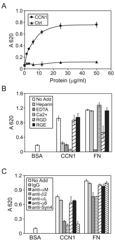
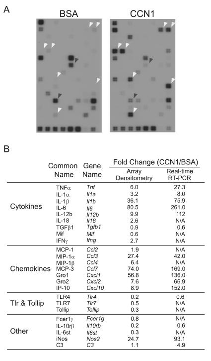
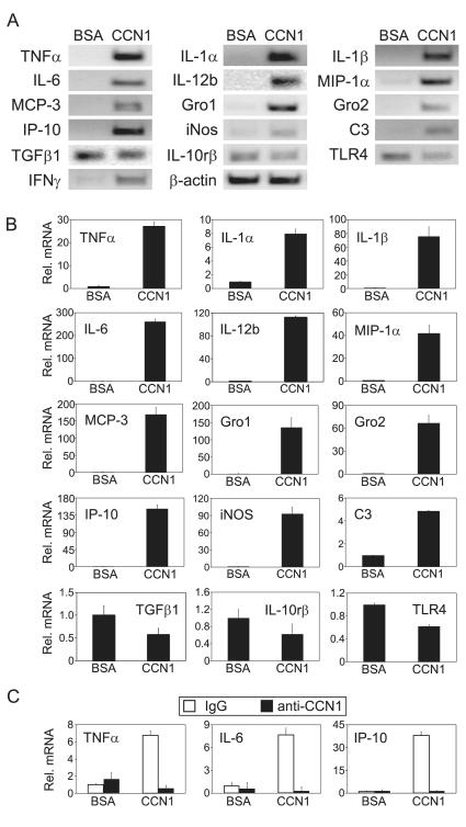
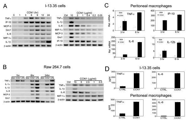
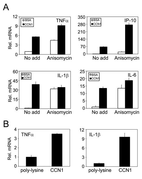
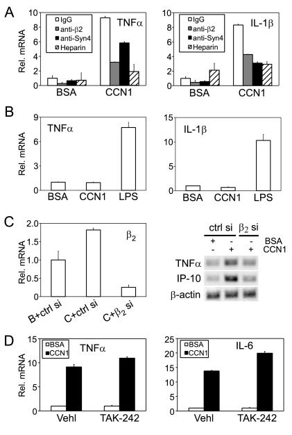
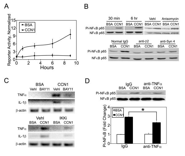
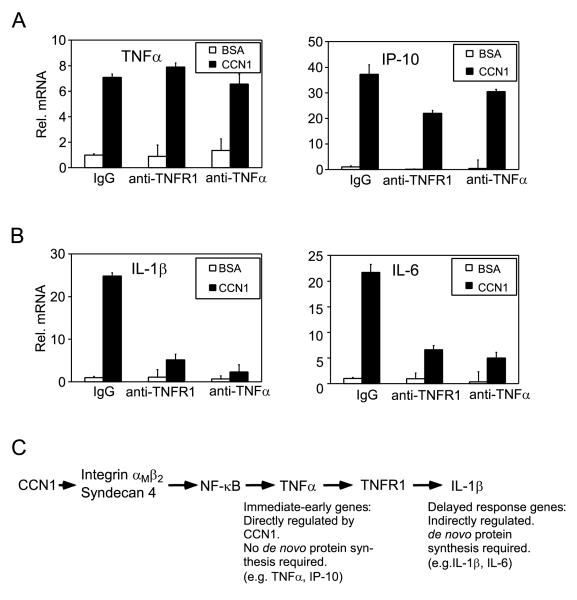
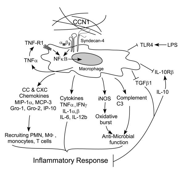
CCN1 (CYR61) is a matricellular protein that is highly expressed at sites of inflammation and wound repair. In these contexts, CCN1 can modify the activities of specific cytokines, enabling TNF-alpha to be cytotoxic without blocking NF-kappaB activity and enhancing the apoptotic activity of Fas ligand and TRAIL. In this paper, we show that CCN1 supports the adhesion of macrophages through integrin alpha(M)beta(2) and syndecan-4, activates NFkappaB-mediated transcription, and induces a proinflammatory genetic program characteristic of classically activated M1 macrophages that participates in Th1 responses. The effects of CCN1 include upregulation of cytokines (TNF-alpha, IL-1alpha, IL-1beta, IL-6, and IL-12b), chemokines (MIP-1alpha; MCP-3; growth-related oncogenes 1 and 2; and inflammatory protein 10), and regulators of oxidative stress and complement (inducible NO synthase and C3) and downregulation of specific receptors (TLR4 and IL-10Rbeta) and anti-inflammatory factors (TGF-beta1). CCN1 regulates this genetic program through at least two distinct mechanisms: an immediate-early response resulting from direct activation of NF-kappaB by CCN1, leading to the synthesis of cytokines including TNF-alpha and inflammatory protein 10; and a delayed response resulting from CCN1-induced TNF-alpha, which acts as an autocrine/paracrine mediator to activate the expression of other cytokines including IL-1beta and IL-6. These results identify CCN1 as a novel component of the extracellular matrix that activates proinflammatory genes in macrophages, implicating its role in regulating macrophage function during inflammation.


Figures

Figure 1
Figure 1. CCN1 supports macrophage adhesion through integrin αMβ2 and syndecan-4
A. Microtiter wells were coated with various concentrations of recombinant CCN1 and control protein (Ctrl) as indicated, onto which I-13.35 macrophages were plated in serum-free medium and incubated at 37 °C for 15 min. Adherent cells were stained with methylene blue and quantified by absorbance at 620 nm. B. Cells were incubated with various inhibitors as indicated (EDTA, 2.5 mM; Ca2+, 5 mM; soluble heparin, 1 μg/ml; GRGDSP and GRGESP peptides, 2 mM each) and cell adhesion assay was performed in dishes coated with CCN1 or fibronectin (FN) as adhesion substrates. C. Cells were pre-incubated with mAbs against integrins αM, αL, α6, β2, or antibodies against syndecan-4, or normal mouse IgG (50 μg/ml each) for 1 hr at room temperature, followed by assay for cell adhesion to CCN1 and FN. Data are mean ± S.D. of triplicate determinations, and all experiments were repeated three times with similar results.
Figure 2
Figure 2. Microarray hybridization
A. Serum-starved I-13.35 macrophages were incubated with 10 μg/ml CCN1 or BSA at 37°C for 6 hr. Total RNA was used to prepare cRNA probes and hybridized to microarrays containing oligodeoxynucleotides corresponding to 113 mouse inflammatory genes. White arrowheads point to representative genes whose expression is elevated in CCN1-treated cells, and black arrowheads point to CCN1 down-regulated genes. B. A list of CCN1 regulated genes. Fold changes were calculated by densitometry scanning of the x-ray films or from results of real-time PCR (see Fig. 3), and was defined as expression levels in CCN1-treated samples divided by BSA-treated controls.
Figure 3
Figure 3. CCN1-regulated inflammatory gene expression
A. Cells were treated with CCN1 or BSA for 6 hr and mRNA levels of indicated genes were analyzed by semi-quantitative RT-PCR. B. Effects of CCN1 on mRNA levels of indicated genes were analyzed by quantitative real-time PCR. mRNA levels in BSA-treated controls were set as one. C. CCN1 protein (10 μg/ml) was incubated with affinity-purified rabbit anti-CCN1 polyclonal antibodies (anti-CCN1) or normal rabbit IgG (100 μg/ml each) at 4°C overnight before being added to cells and incubated for 3 hrs. The mRNA levels of indicated genes were analyzed by quantitative real-time PCR.
Figure 4
Figure 4. Time course and dosage dependence of CCN1-regulated inflammatory gene expression
CCN1 regulated gene expression was tested in two macrophage cell lines: I-13.35 (panels in A), and Raw 264.7 (panels in B). Serum-starved cells were incubated with CCN1 (10 μg/ml) for various times from 0 ~ 24 hr as indicated (left panels in A, B), or cells were treated with various concentrations of CCN1 as indicated for 6 hr, and gene expression was assessed using semi-quantitative RT-PCR. C. Expression of TNFα, IP-10, IL-6, and IL-12b in freshly isolated peritoneal macrophages from wild type C57BL/6 mice treated with BSA or CCN1 for 3 or 6 hrs was assayed by real-time RT-PCR. D. The amount of TNFα and IL-6 proteins detected in I-13.35 cells or conditioned medium, respectively (upper panel) and in conditioned medium of freshly isolated peritoneal macrophages (lower panel) was detected by ELISA.
Figure 5
Figure 5. Characterization of CCN1-regulated inflammatory gene expression
A. Cells were incubated with CCN1 protein either with or without anisomycin (10 μM) and treated with BSA or CCN1 for 6 hrs. Relative levels of TNFα, IP-10, IL-1β, and IL-6 mRNA were analyzed by real-time RT-PCR. B. Cells were adhered for 3 hr on culture dishes coated with CCN1 (10 μg/ml) or the inert substrate poly-L-lysine (5 μg/ml). Relative TNFα and IL-1β mRNAs levels were determined by real-time RT-PCR.
Figure 6
Figure 6. Integrin β2 and syndecan-4, but not TLR4, are critical for CCN1-regulated gene expression
A. I-13.35 cells were pre-incubated for 30 min with an anti-β2 mAb (100 μg/ml), anti-syndecan-4 antibodies (anti-Syn 4, 40 μg/ml) or nonspecific IgG (100 μg/ml) before being treated with CCN1 for 6 hr. Where indicated, cells were treated with CCN1 in the presence of soluble heparin (2 μg/ml) in the culture media. The levels of TNFα and IL-1β mRNA were analyzed by real-time RT-PCR. B. Peritoneal macrophages isolated from integrin αM knock-out mice were treated for 6 hrs with CCN1 or LPS. Relative levels of TNFα and IL-1β mRNA were analyzed by real-time RT-PCR. C. I-13.35 cells were transfected with siRNA against integrin β22 si) or a control non-targeting siRNA (ctrl si) for 48 hrs. Cells were then incubated with CCN1 or BSA for 3 hrs. Down-regulation of β2 mRNA was assessed by real-time RT-PCR (left panel). Expression of TNFα and IP-10 was evaluated by semi-quantitative RT-PCR. D. Cells were incubated with the TLR4 inhibitor TAK-242 (0.5 μM) or vehicle DMSO (Vehl) for 30 min. prior to CCN1 treatment. TNFα and IL-1β mRNA levels were measured by real-time RT-PCR.
Figure 7
Figure 7. CCN1 activates NFκB in macrophages
A. I-13.35 cells were co-transfected the luciferase reporter pNFκB-Luc driven by multiple NFκB-responsive elements (57), and pRL-CMV, a construct for transfection efficiency controls. Cells were treated with CCN1 or BSA one day later for the indicated times, and luciferase activity in cell lysates determined and normalized against transfection efficiency controls. B. NFκB activation was assessed by immunoblotting of cell lysates resolved on SDS-PAGE with antibodies against total and phosphorylated NFκB-p65. Upper panel: cells were treated with CCN1 for 30 min. or 6 hrs, or for 3 hrs with and without anisomycin (10 μM) or its vehicle DMSO. Lower panel, cells were preincubated with antibodies against integrin β2 (100 μg/ml) or syndecan-4 (40 μg/ml) for 30 min. before being treated with CCN1 for 1.5 hr. C. Cells were pre-incubated with NFκB signaling inhibitors for 30 min (BAY11, 5 μM IκB complex inhibitor BAY11-7082; IKKi, 50 μM IκB kinase inhibitor) before being treated with CCN1. TNFα and IL-1β mRNA expression was analyzed by semi quantitative RT-PCR. D. Cells were incubated with anti-TNFα (100 μg/ml) antibodies or normal IgG for 30 min and treated with CCN1 for 6 hrs. Phosphorylation on NFκB-p65 was analyzed by immunoblotting and quantified by densitometry. *: p<0.05.
Figure 8
Figure 8. A role of CCN1-induced TNFα as mediator for IL-1β and IL-6 induction
Macrophages were pre-incubated for 30 min with a monoclonal antibody blocking TNF-R1, ployclonal antibodies neutralizing TNFα, or a normal IgG (100 μg/ml each), before being treated with CCN1 for 6 hr. and total RNA isolated for gene expression study using real-time RT-PCR. Expression of TNFα and IP-10 (A), and of IL-1β and IL-6 (B) is shown. C. CCN1 activates NFκB (Fig. 7A, B) through β2 integrin and syndecan-4 (Fig. 6), leading to the expression of NFκB-inducible genes such as TNFα and IP-10. Other genes, exemplified by IL-1β, are upregulated through de novo protein synthesis (Fig. 5A) of CCN1-induced TNFα (Fig. 8B).
Figure 9
Figure 9. A model for CCN1 regulation of inflammatory genes in macrophages
The matricellular protein CCN1 induces gene expression changes characteristic of the classically activated M1-type response in macrophages through its cell surface receptors integrin αMβ2 and syndecan-4, resulting in elevated expression of pro-inflammatory cytokines like TNFα, IL-1β, IL-6, and IFNγ, and chemokines including MIP-1α, MCP-3, Gro1, Gro2, and IP-10. These factors may recruit and activate more inflammatory cells including polymorphonuclear neutrophils, monocyte/macrophages, and T-lymphocytes. Simultaneously, expression of anti-inflammatory effectors such as TGF-β1 and IL-10rβ are attenuated, resulting in a further amplification of inflammatory response. CCN1 may enhance host defense against invading bacteria by upregulating macrophage expressions of iNOS, which is critical for suppressing bacterial replication (58), and complement C3 protein, which is required for activation of the complement system. On the other hand, CCN1 may also confine the host response to invading microbes by down-regulating the LPS receptor TLR4. Collectively, our data support the hypothesis that CCN1 exerts strong pro-inflammation response on macrophages by promoting diverse inflammatory gene expressions.

Similar articles

Cited by

References

    1. Aszodi A, Legate KR, Nakchbandi I, Fassler R. What mouse mutants teach us about extracellular matrix function. Annu. Rev. Cell Dev. Biol. 2006;22:591. - PubMed
    1. Adair-Kirk TL, Senior RM. Fragments of extracellular matrix as mediators of inflammation. Int. J. Biochem. Cell Biol. 2008;40:1101. - PMC - PubMed
    1. O’Reilly PJ, Gaggar A, Blalock JE. Interfering with extracellular matrix degradation to blunt inflammation. Curr. Opin. Pharmacol. 2008;8:242. - PMC - PubMed
    1. Bornstein P, Sage EH. Matricellular proteins: extracellular modulators of cell function. Curr. Opin. Cell Biol. 2002;14:608. - PubMed
    1. Scatena M, Liaw L, Giachelli CM. Osteopontin: a multifunctional molecule regulating chronic inflammation and vascular disease. Arterioscler. Thromb. Vasc. Biol. 2007;27:2302. - PubMed

Publication types

MeSH terms

Associated data