. 2010 Mar 3;102(5):340-51.
doi: 10.1093/jnci/djp535. Epub 2010 Feb 17.

Prognostic significance of tumorigenic cells with mesenchymal features in pancreatic adenocarcinoma

Affiliations

Prognostic significance of tumorigenic cells with mesenchymal features in pancreatic adenocarcinoma

Zeshaan A Rasheed et al. J Natl Cancer Inst. .

Abstract

Background: Specific populations of highly tumorigenic cells are thought to exist in many human tumors, including pancreatic adenocarcinoma. However, the clinical significance of these tumor-initiating (ie, cancer stem) cells remains unclear. Aldehyde dehydrogenase (ALDH) activity can identify tumor-initiating cells and normal stem cells from several human tissues. We examined the prognostic significance and functional features of ALDH expression in pancreatic adenocarcinoma.

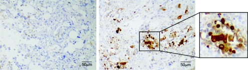
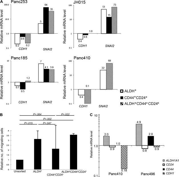
Methods: ALDH expression was analyzed by immunohistochemistry in 269 primary surgical specimens of pancreatic adenocarcinoma and examined for association with clinical outcomes and in paired primary tumors and metastatic lesions from eight pancreatic cancer patients who had participated in a rapid autopsy program. The clonogenic growth potential of ALDH-positive pancreatic adenocarcinoma cells was assessed in vitro by a colony formation assay and by tumor growth in immunodeficient mice (10-14 mice per group). Mesenchymal features of ALDH-positive pancreatic tumor cells were examined by using quantitative reverse transcription-polymerase chain reaction and an in vitro cell invasion assay. Gene expression levels and the invasive potential of ADLH-positive pancreatic cancer cells relative to the bulk cell population were examined by reverse transcription-polymerase chain reaction and an in vitro invasion assays, respectively. All statistical tests were two-sided.

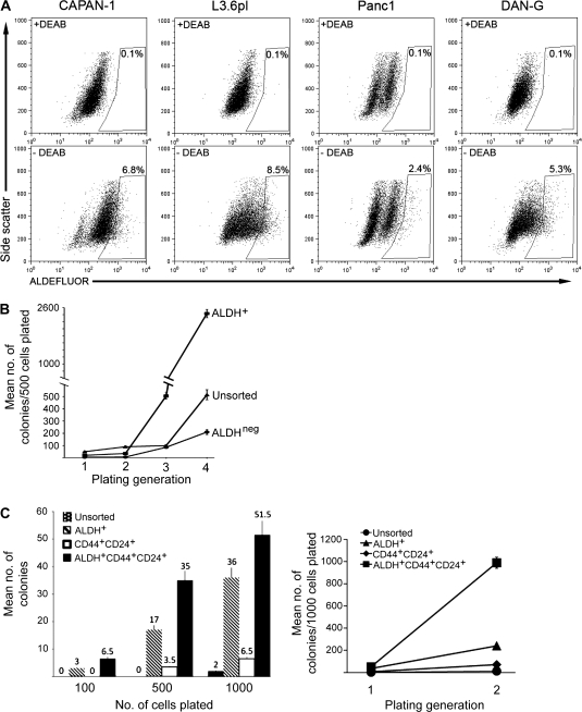
Results: ALDH-positive tumor cells were detected in 90 of the 269 primary surgical specimens, and their presence was associated with worse survival (median survival for patients with ALDH-positive vs ALDH-negative tumors: 14 vs 18 months, hazard ratio of death = 1.28, 95% confidence interval = 1.02 to 1.68, P = .05). Six (75%) of the eight patients with matched primary and metastatic tumor samples had ALDH-negative primary tumors, and in four (67%) of these six patients, the matched metastatic lesions (located in liver and lung) contained ALDH-positive cells. ALDH-positive cells were approximately five- to 11-fold more clonogenic in vitro and in vivo compared with unsorted or ALHD-negative cells, expressed genes consistent with a mesenchymal state, and had in vitro migratory and invasive potentials that were threefold greater than those of unsorted cells.

Conclusions: ALDH expression marks pancreatic cancer cells that have stem cell and mesenchymal features. The enhanced clonogenic growth and migratory properties of ALDH-positive pancreatic cancer cells suggest that they play a key role in the development of metastatic disease that negatively affects the overall survival of patients with pancreatic adenocarcinoma.


Figures

Figure 1
Figure 1
Association between aldehyde dehydrogenase (ALDH) expression in primary pancreatic tumors and patient survival. A) Immunohistochemical localization of ALDH protein expression in pancreatic intraepithelial neoplastic lesions. Representative images show staining for ALDH (brown) in a high-grade pancreatic intraepithelial neoplastic lesion (ie, PanIN-3 or carcinoma in situ; right panel and inset) and in a low-grade pancreatic intraepithelial neoplastic lesion (ie, PanIN-2; left panel). B) Immunohistochemical staining of ALDH in primary human pancreatic adenocarcinoma. Left panel shows an ALDH-positive tumor in which intensely staining cells are found mostly at the base of cancerous glands (arrowheads). Right panel shows an ALDH-negative tumor. C) Kaplan–Meier analysis of overall survival for patients who underwent pancreatectomy based on tumor ALDH expression by immunohistochemistry. At 24 months, the number of patients at risk with ALDH-positive and ALDH-negative tumors was 27 (proportion surviving = 0.30, 95% confidence interval [CI] = 0.22 to 0.42) and 67 (proportion surviving = 0.39, 95% CI = 0.32 to 0.47), respectively. At 48 months, the number of patients at risk with ALDH-positive and ALDH-negative tumors was 9 (proportion surviving = 0.15, 95% CI = 0.09 to 0.25) and 33 (proportion surviving = 0.23, 95% CI = 0.18 to 0.31), respectively. At 72 months, the number of patients at risk with ALDH-positive and ALDH-negative tumors was 2 (proportion surviving = 0.11, 95% CI = 0.05 to 0.21) and 16 (proportion surviving = 0.17, 95% CI = 0.12 to 0.24).
Figure 2
Figure 2
Cancer stem cell properties of aldehyde dehydrogenase (ALDH)–positive pancreatic cancer cells. A) Flow cytometry. ALDH activity in human pancreatic cancer cell lines CAPAN-1, DAN-G, Panc1, and L3.6pl was measured by flow cytometry by using the ALDEFLUOR reagent in the presence (top row) and absence (bottom row) of the ALDH1 inhibitor diethylamino-benzaldehyde (DEAB). The frames represent gates that depict ALDH-positive (ALDH+) cells that were created based on the flow cytometry plot from cells treated with DEAB and then applied to the plots of untreated cells. The percentages of ALDH+ cells are shown in or above the gate. B) Colony formation assay. ALDH+, ALDH-negative (ALDHneg), and unsorted CAPAN-1 cells were isolated by fluorescence-activated cell sorting and evaluated for clonogenic growth in vitro. Five hundred cells were plated in methylcellulose, and colonies were counted 10 days later. Serial replating was carried out for four generations. Error bars correspond to 95% confidence intervals. C) Colony formation assay with serially diluted cells. Unsorted, ALDH+, CD44- and CD24-positive (CD44+CD24+), and ALDH-, CD24-, and CD44-positive (ALDH+CD44+CD24+) CAPAN-1 cells were isolated by fluorescence-activated cell sorting and evaluated for clonogenic growth as described above. The left panel depicts the number of colonies that were counted after varying numbers of cells (100, 500, or 1000) were plated in methylcellulose. The right panel depicts the number of colonies that grew after 1000 cells were plated in methylcellulose and serially replated for a second generation. Mean values (n = 3) with 95% confidence intervals (error bars) are depicted.
Figure 3
Figure 3
Aldehyde dehydrogenase (ALDH) expression in a primary tumor and metastatic lesion from a pancreatic cancer patient who participated in the rapid autopsy program. Immunohistochemical staining for ALDH (brown) in the primary tumor (left panel) and in a lung metastasis (right panel).
Figure 4
Figure 4
Properties of pancreatic cancer stem cells (CSCs). A) E-cadherin and SLUG mRNA expression in pancreatic CSC. Unsorted, ALDH-positive (ALDH+), CD44- and CD24-positive (CD44+CD24+), and ALDH-, CD24-, and CD44-positive (ALDH+CD44+CD24+) cells from four human xenografts (Panc253, Panc185, JH015, and Panc410) were isolated by fluorescence-activated cell sorting and analyzed for ACTB (β-actin), CDH1, and SNAI2 mRNA levels by real-time quantitative reverse transcription–polymerase chain reaction (RT-PCR) in triplicate. Results are depicted as normalized gene expression in ALDH+, CD44+CD24+, and ALDH+CD44+CD24+ cells relative to unsorted cells. One experiment was performed for each xenograft. B) Cell migration assay. Unsorted, ALDH+, CD44+CD24+, and ALDH+CD44+CD24+ CAPAN-1 cells were isolated by fluorescence-activated cell sorting and applied to a transwell chambers containing filters with 8-μm pores. Cells that had migrated through the filter were counted 72 hours later. The relative number of migrating cells from each cell population compared with the number of migrating cells from the unsorted population is plotted. The results plotted are from three separate experiments, each containing duplicate wells. Error bars correspond to 95% confidence intervals. P values (two-sided) are from a Student t test. C) Cell invasion assay. Low-passage human pancreatic cancer xenografts from mice (Panc410 and Panc496) were made into single-cell suspensions, depleted of mouse cells by using an LD cell separation column, applied to a Matrigel-coated invasion chamber, and incubated for 72 hours. ALDH1A1, CD44, and CD24 mRNA levels in the invasive and noninvasive cells were measured by quantitative RT-PCR in triplicate. Results are depicted as normalized gene expression in invasive cells relative to noninvasive cells. One experiment was performed for each xenograft.

References

    1. Jemal A, Siegel R, Ward E, et al. Cancer statistics, 2008. CA Cancer J Clin. 2008;58(2):71–96. - PubMed
    1. Sohn TA, Yeo CJ, Cameron JL, et al. Resected adenocarcinoma of the pancreas-616 patients: results, outcomes, and prognostic indicators. J Gastrointest Surg. 2000;4(6):567–579. - PubMed
    1. Winter JM, Cameron JL, Campbell KA, et al. 1423 pancreaticoduodenectomies for pancreatic cancer: a single-institution experience. J Gastrointest Surg. 2006;10(9):1199–1210. discussion 1210–1211. - PubMed
    1. Hermann PC, Huber SL, Herrler T, et al. Distinct populations of cancer stem cells determine tumor growth and metastatic activity in human pancreatic cancer. Cell Stem Cell. 2007;1(3):313–323. - PubMed
    1. Li C, Heidt DG, Dalerba P, et al. Identification of pancreatic cancer stem cells. Cancer Res. 2007;67(3):1030–1037. - PubMed

Publication types

MeSH terms

Substances