. 2010 Jul;51(7):3485-93.
doi: 10.1167/iovs.09-5120. Epub 2010 Feb 17.

Effects of intracranial trochlear neurectomy on the structure of the primate superior oblique muscle

Affiliations

Effects of intracranial trochlear neurectomy on the structure of the primate superior oblique muscle

Joseph L Demer et al. Invest Ophthalmol Vis Sci. 2010 Jul.

Abstract

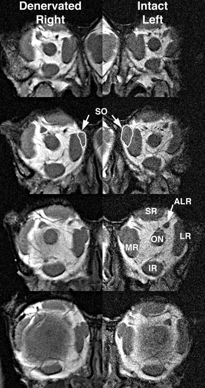
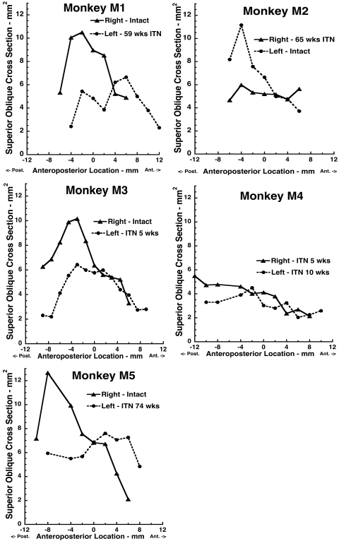
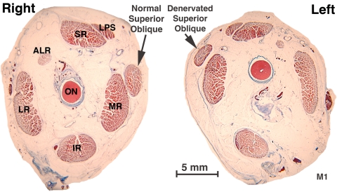
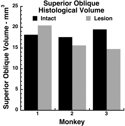
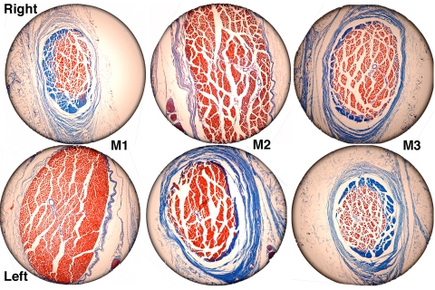
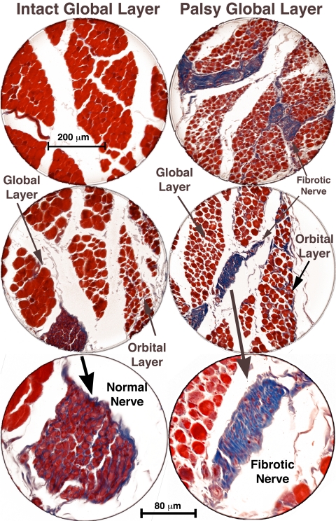
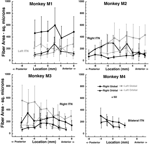
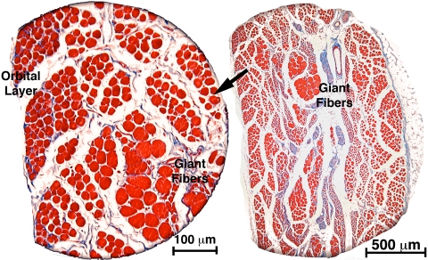
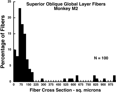
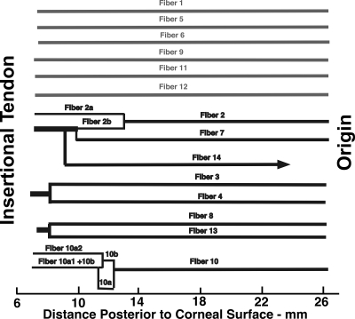
PURPOSE. Although cyclovertical strabismus in humans is frequently attributed to superior oblique (SO) palsy, anatomic effects of SO denervation have not been studied. Magnetic resonance imaging (MRI) and orbital histology was used to study the effects of acute trochlear (CN4) denervation on the monkey SO. METHODS. Five juvenile macaque monkeys were perfused with formalin for 5 weeks: 15 months after unilateral or bilateral 10-mm intracranial trochlear neurectomy. Denervated and fellow orbits were imaged by MRI, embedded whole in paraffin, serially sectioned at 10-mum thickness, and stained with Masson trichrome. Whole muscle and individual fiber cross sections were quantified in SO muscles throughout the orbit and traced larger fibers in one specimen where they were present. RESULTS. MRI demonstrated marked reduction in midorbital cross section in denervated SO muscles, with anterior shift of SO mass preserving overall volume. Muscle fibers exhibited variable atrophy along their lengths. Denervated orbital layer (OL) fiber cross sections were slightly but significantly reduced from control at most anteroposterior locations, but this reduction was much more profound in global layer (GL) fibers. Intraorbital and intramuscular CN4 were uniformly fibrotic. In one animal, there were scattered clusters of markedly hypertrophic GL fibers that exhibited only sparse myomyous junctions only anteriorly. CONCLUSIONS. CN4 denervation produces predominantly SO GL atrophy with relative OL sparing. Overall midorbital SO atrophy was evident by MRI as early as 5 weeks after denervation, as denervated SO volume shifted anteriorly. Occasional GL fiber hypertrophy suggests that at least some SO fibers extend essentially the full muscle length after trochlear neurectomy.


Figures

Figure 1.
Figure 1.
MRI of both orbits of monkey M2 in 2-mm-thick quasi-coronal planes spaced at 4-mm intervals from posterior (top) to anterior (bottom). Note smaller SO muscle (white outline) in the deep orbit on the right side denervated by ITN. ALR, accessory lateral rectus muscle; IR, inferior rectus muscle; LR, lateral rectus muscle; MR, medial rectus muscle; ON, optic nerve; SO, superior oblique muscle; SR, superior rectus muscle.
Figure 2.
Figure 2.
MRI determinations of SO cross sections in monkeys M1 to M5 as functions of anteroposterior location relative to the junction of the globe with the optic nerve, designated as 0. All denervated SO muscles exhibited reduced cross-sectional area posterior to the globe.
Figure 3.
Figure 3.
Coronal histologic sections of the right and left orbits of monkey M1, 56 weeks after left ITN, at the globe–optic nerve junction, designated as image plane 0, showing left SO muscle atrophy. Masson trichrome. Abbreviations as in Figure 1.
Figure 4.
Figure 4.
Total gross volumes of SO muscles determined from coronal histologic sections in monkeys M1 to M3 who had undergone unilateral ITN. Note the absence of consistent volume reduction by ITN.
Figure 5.
Figure 5.
Coronal histologic sections of the SO taken 8 mm anterior to the globe–optic nerve junction showing anterior shift of fibers in the denervated left orbit of monkey M1 and the denervated right orbits of monkeys M2 and M3. Masson trichrome. Circular field diameter, 2.2 mm.
Figure 6.
Figure 6.
Coronal histologic sections of the right and left orbits of monkey M1, 56 weeks after left ITN, showing marked atrophy of global layer fibers in the left SO muscle, with preservation of left orbital layer fibers. Fibrotic intramuscular nerves (shown at higher power in bottom panels) in the palsied left SO stain blue with Masson trichrome, whereas intact nerves in the right SO stain purple. Scale bars: (top two panels) 200 μm; (bottom panel) 80 μm.
Figure 7.
Figure 7.
Mean cross sectional areas of fibers in the SO GL and OL. Monkey M1 had undergone left ITN, M2 and M3 underwent right ITN, and M4 underwent bilateral ITN, but data are analyzed only for the right side. Note the marked reduction in denervated GL, but not OL fiber cross section. Each data point is the mean of 50 fibers. The abscissa is aligned on the globe–optic nerve junction.
Figure 8.
Figure 8.
Coronal histologic section of the right orbit of monkey M2, 60 weeks after right ITN, showing clusters of giant fibers among the severely atrophic fibers in the GL. Masson trichrome stain. Section taken 2 mm posterior to the globe–optic nerve junction.
Figure 9.
Figure 9.
Histogram of GL fiber cross sections in the right orbit of monkey M2, 60 weeks after right ITN, showing clusters of giant GL fibers among the severely atrophic fibers in the GL. Mode for atrophic fibers is 75 μm2, but some hypertropic fibers exhibited cross sections of up to 1000 μm2 so that the mean for all fibers was 158 μm2. Section were taken 2 mm posterior to the globe–optic nerve junction and are illustrated in Figure 8.
Figure 10.
Figure 10.
Longitudinal tracing of 14 identified giant SO GL fibers in the right orbit of monkey M2 after right ITN. Six fibers extended from origin to tendon without myomyous junctions, whereas eight fibers exhibited anterior myomyous junctions.

Similar articles

Cited by

References

    1. Bielschowsky A. Lectures on motor anomalies. XI. Etiology, prognosis, and treatment of ocular paralyses. Am J Ophthalmol 1939;22:723–734
    1. von Noorden GK, Murray E, Wong SY. Superior oblique paralysis: a review of 270 cases. Arch Ophthalmol 1986;104:1771–1776 - PubMed
    1. Adler FE. Physiologic factors in differential diagnosis of paralysis of superior rectus and superior oblique muscles. Arch Ophthalmol 1946;36:661–673 - PubMed
    1. Scott WE, Kraft SP. Classification and treatment of superior oblique palsies: II. Bilateral superior oblique palsies. In: Caldwell D. ed. Pediatric Ophthalmology and Strabismus: Transactions of the New Orleans Academy of Ophthalmology New York: Raven Press; 1986:265–291 - PubMed
    1. Scott WE, Parks MM. Differential diagnosis of vertical muscle palsies. In: von Noorden GK. ed. Symposium on Strabismus: Transactions of the New Orleans Academy of Ophthalmology.St. Louis: Mosby; 1978:118–134

Publication types