. 2010 Jul;51(7):3687-700.
doi: 10.1167/iovs.09-4766. Epub 2010 Feb 17.

Spectrum of rhodopsin mutations in French autosomal dominant rod-cone dystrophy patients

Affiliations

Spectrum of rhodopsin mutations in French autosomal dominant rod-cone dystrophy patients

Isabelle Audo et al. Invest Ophthalmol Vis Sci. 2010 Jul.

Abstract

PURPOSE. To identify the prevalence of rhodopsin (RHO) mutations in French patients with autosomal dominant rod-cone dystrophies (adRPs). Methods. Detailed phenotypic characterization was performed, including precise family history, best corrected visual acuity with the ETDRS chart, slit lamp examination, kinetic and static perimetry, full-field and multifocal electroretinography (ERG), fundus autofluorescence imaging (FAF), and optical coherence tomography (OCT). For genetic diagnosis, genomic DNA of 79 families was isolated by standard

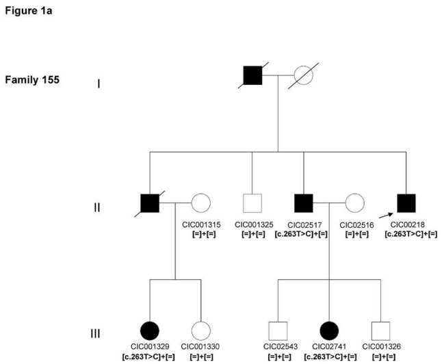
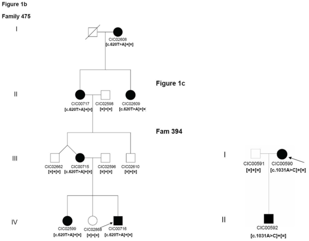
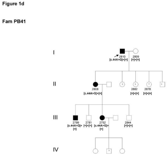
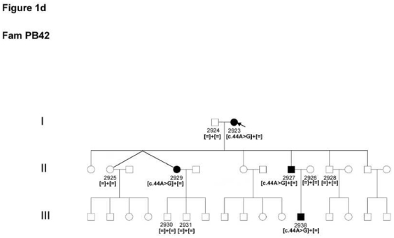
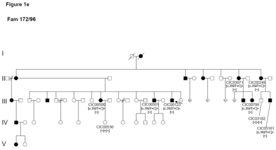
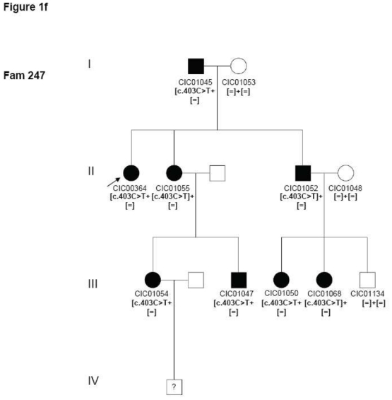
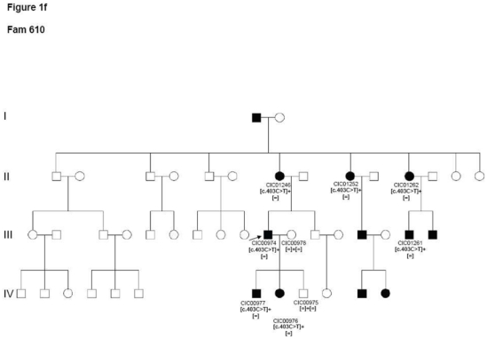
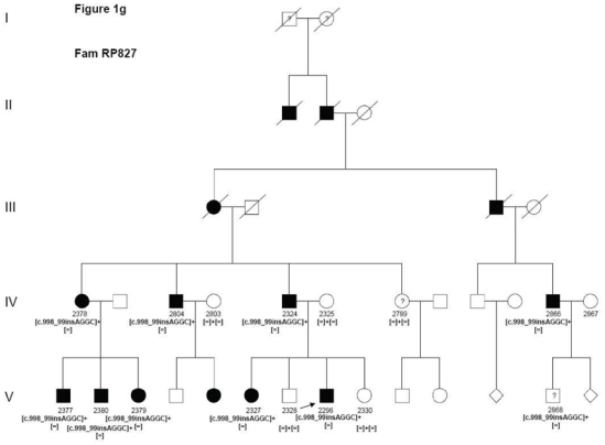
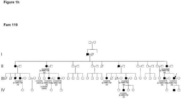
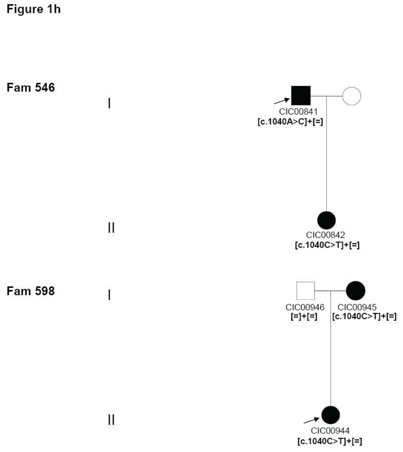
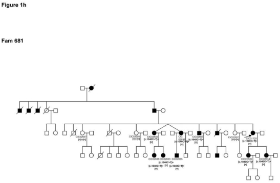
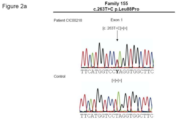
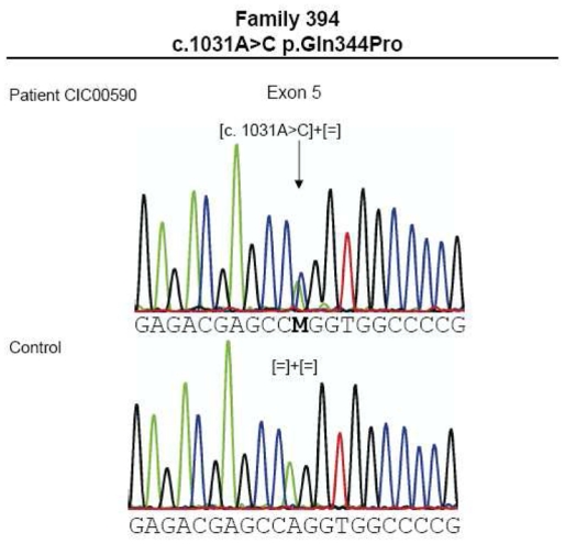
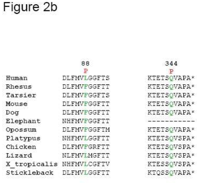
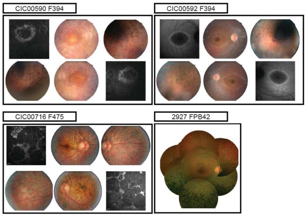
Methods: The coding exons and flanking intronic regions of RHO were PCR amplified, purified, and sequenced in the index patient. RESULTS. Of this French adRP sample, 16.5% carried an RHO mutation. Three different families showed a novel mutation (p. Leu88Pro, p.Met207Lys and p.Gln344Pro), while ten unrelated families showed recurrent, previously published mutations (p.Asn15Ser, p.Leu131Pro, p.Arg135Trp, p.Ser334GlyfsX21 and p.Pro347Leu). All mutations co-segregated with the phenotype within a family, and the novel mutations were not identified in control samples. CONCLUSIONS. This study revealed that the prevalence of RHO mutations in French adRP patients is in accordance with that in other studies from Europe. Most of the changes identified herein reflect recurrent mutations, within which p.Pro347Leu substitution is the most prevalent. Nevertheless, almost one fourth of the changes are novel, indicating that, although RHO is the first gene implicated and probably the most studied gene in RP, it is still important performing mutation analysis in RHO to detect novel changes. The detailed phenotype-genotype analyses in all available family members deliver the basis for therapeutic approaches in those families.


Figures

Figure 1
Figure 1
Pedigrees of adRP patients with RHO mutations and co-segregation in available family members. Filled symbols represent affected and unfilled unaffected persons. Squares indicate males, circles females. Arrows reflect the index patients.
Figure 1
Figure 1
Pedigrees of adRP patients with RHO mutations and co-segregation in available family members. Filled symbols represent affected and unfilled unaffected persons. Squares indicate males, circles females. Arrows reflect the index patients.
Figure 1
Figure 1
Pedigrees of adRP patients with RHO mutations and co-segregation in available family members. Filled symbols represent affected and unfilled unaffected persons. Squares indicate males, circles females. Arrows reflect the index patients.
Figure 1
Figure 1
Pedigrees of adRP patients with RHO mutations and co-segregation in available family members. Filled symbols represent affected and unfilled unaffected persons. Squares indicate males, circles females. Arrows reflect the index patients.
Figure 1
Figure 1
Pedigrees of adRP patients with RHO mutations and co-segregation in available family members. Filled symbols represent affected and unfilled unaffected persons. Squares indicate males, circles females. Arrows reflect the index patients.
Figure 1
Figure 1
Pedigrees of adRP patients with RHO mutations and co-segregation in available family members. Filled symbols represent affected and unfilled unaffected persons. Squares indicate males, circles females. Arrows reflect the index patients.
Figure 1
Figure 1
Pedigrees of adRP patients with RHO mutations and co-segregation in available family members. Filled symbols represent affected and unfilled unaffected persons. Squares indicate males, circles females. Arrows reflect the index patients.
Figure 1
Figure 1
Pedigrees of adRP patients with RHO mutations and co-segregation in available family members. Filled symbols represent affected and unfilled unaffected persons. Squares indicate males, circles females. Arrows reflect the index patients.
Figure 1
Figure 1
Pedigrees of adRP patients with RHO mutations and co-segregation in available family members. Filled symbols represent affected and unfilled unaffected persons. Squares indicate males, circles females. Arrows reflect the index patients.
Figure 1
Figure 1
Pedigrees of adRP patients with RHO mutations and co-segregation in available family members. Filled symbols represent affected and unfilled unaffected persons. Squares indicate males, circles females. Arrows reflect the index patients.
Figure 1
Figure 1
Pedigrees of adRP patients with RHO mutations and co-segregation in available family members. Filled symbols represent affected and unfilled unaffected persons. Squares indicate males, circles females. Arrows reflect the index patients.
Figure 2
Figure 2
(a). Electropherograms of novel RHO mutations highlighted by an arrow. (b) Multiple amino acid sequence alignments of different species of novel mutated residues (depicted in green). Amino acid substitutions are highlighted in red. The position of the respective amino acids is shown in black numbers.
Figure 2
Figure 2
(a). Electropherograms of novel RHO mutations highlighted by an arrow. (b) Multiple amino acid sequence alignments of different species of novel mutated residues (depicted in green). Amino acid substitutions are highlighted in red. The position of the respective amino acids is shown in black numbers.
Figure 3
Figure 3
Fundus and autofluorescence pictures of 3 index patients with distinct adRP phenotypes (diffuse, sector RP and restricted chorioretinal atrophy)

Similar articles

Cited by

References

    1. Hartong DT, Berson EL, Dryja TP. Retinitis pigmentosa. Lancet. 2006;368:1795–1809. - PubMed
    1. Dryja TP, McEvoy JA, McGee TL, Berson EL. Novel rhodopsin mutations Gly114Val and Gln184Pro in dominant retinitis pigmentosa. Invest Ophthalmol Vis Sci. 2000;41:3124–3127. - PubMed
    1. Dryja TP, Hahn LB, Cowley GS, McGee TL, Berson EL. Mutation spectrum of the rhodopsin gene among patients with autosomal dominant retinitis pigmentosa. Proc Natl Acad Sci U S A. 1991;88:9370–9374. - PMC - PubMed
    1. Sung CH, Davenport CM, Hennessey JC, et al. Rhodopsin mutations in autosomal dominant retinitis pigmentosa. Proc Natl Acad Sci U S A. 1991;88:6481–6485. - PMC - PubMed
    1. Macke JP, Davenport CM, Jacobson SG, et al. Identification of novel rhodopsin mutations responsible for retinitis pigmentosa: implications for the structure and function of rhodopsin. Am J Hum Genet. 1993;53:80–89. - PMC - PubMed

MeSH terms