Review
. 2010;20(2):369-93.
doi: 10.3233/JAD-2010-1375.

Oxidatively modified glyceraldehyde-3-phosphate dehydrogenase (GAPDH) and Alzheimer's disease: many pathways to neurodegeneration

Affiliations
Review

Oxidatively modified glyceraldehyde-3-phosphate dehydrogenase (GAPDH) and Alzheimer's disease: many pathways to neurodegeneration

D Allan Butterfield et al. J Alzheimers Dis. 2010.

Abstract

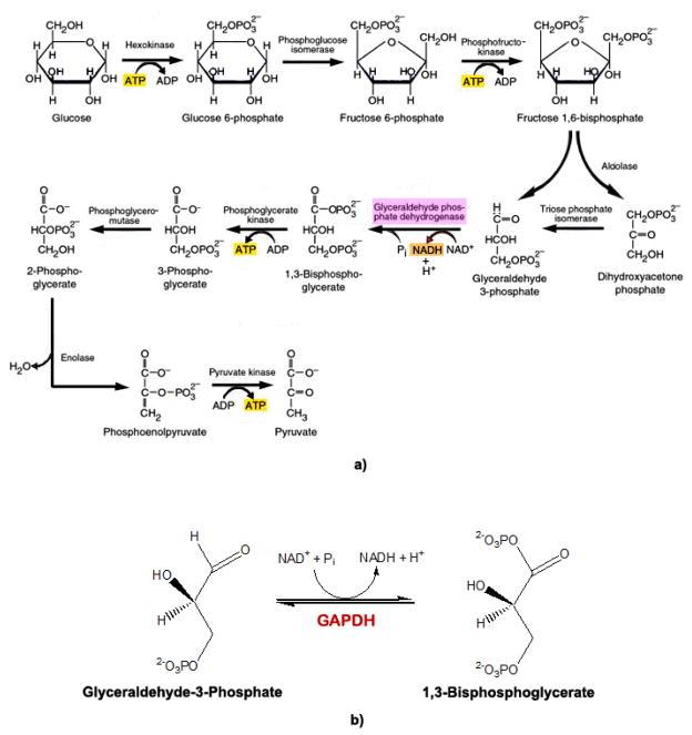
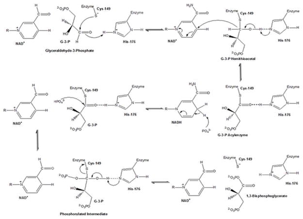
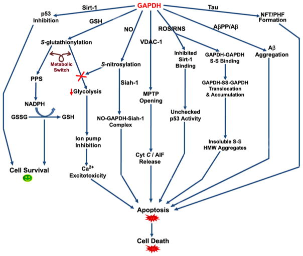
Recently, the oxidoreductase, glyceraldehyde-3-phosphate dehydrogenase (GAPDH), has become a subject of interest as more and more studies reveal a surfeit of diverse GAPDH functions, extending beyond traditional aerobic metabolism of glucose. As a result of multiple isoforms and cellular locales, GAPDH is able to come in contact with a variety of small molecules, proteins, membranes, etc., that play important roles in normal and pathologic cell function. Specifically, GAPDH has been shown to interact with neurodegenerative disease-associated proteins, including the amyloid-beta protein precursor (AbetaPP). Studies from our laboratory have shown significant inhibition of GAPDH dehydrogenase activity in Alzheimer's disease (AD) brain due to oxidative modification. Although oxidative stress and damage is a common phenomenon in the AD brain, it would seem that inhibition of glycolytic enzyme activity is merely one avenue in which AD pathology affects neuronal cell development and survival, as oxidative modification can also impart a toxic gain-of-function to many proteins, including GAPDH. In this review, we examine the many functions of GAPDH with respect to AD brain; in particular, the apparent role(s) of GAPDH in AD-related apoptotic cell death is emphasized.


Figures

Figure 1
Figure 1. GAPDH Structure
Ribbon view of the human placental GAPDH, subunits O, P, Q, and R. GAPDH active-sites clefts are indicated by arrows on each of the asymmetric subunits, P, Q, and R. Color codes: Green, subunit Q; Yellow, subunit O; Blue, subunit P; Red, subunit R; Gray, backbone carbon loops connecting secondary structure successions; Gray molecular structures, NAD(+). Data were obtained from the RCSB protein data bank as entry code 1u8f.pdb, DOI: 10.1107/S0907444905042289 [2]. This figure was drawn using the DeepView Swiss-PDB viewer program, version 4.0.
Figure 2
Figure 2. GAPDH active-site
a) View of Bacillus stearothermophilus GAPDH active-site catalytic residues with a Cys149Ser substitution and occupied by D-G3P and NAD(+). Residues shown are those directly involved in G3P binding (human amino acid analogs discussed in Section 2.0). The structure was drawn in CPK format: Gray, carbons atoms; Blue, nitrogen atoms; Red, oxygen atoms; Orange, phosphorous atoms. Dotted lines represent potential electrostatic or non-polar interactions between G3P, NAD(+), and amino acids; residues T208 and Y311 are normally directly involved with binding of the C149 sulfate anion, however, the C149S substitution replaces the SH-group with a hydroxyl, as shown. Data were obtained from the RCSB protein data bank as entry code 1NQO.pdb, DOI: 10.1074/jbc.M211040200 [77]. This figure was drawn using the DeepView Swiss-PDB viewer program, version 4.0. b) View of human placental GAPDH active-site catalytic residues with no substitution and occupied only by NAD(+). Residues shown are those directly involved in NAD+ binding. The structure was drawn in CPK format: Gray, carbons atoms; Blue, nitrogen atoms; Red, oxygen atoms; Orange, phosphorous atoms; Yellow, sulfur atoms. Dotted lines represent potential electrostatic or non-polar interactions between NAD(+) and specified amino acids. Data were obtained from the RCSB protein data bank as entry code 1u8f.pdb, DOI: 10.1107/S0907444905042289 [2]. This figure was drawn using the DeepView Swiss-PDB viewer program, version 4.0.
Figure 3
Figure 3
a) Glycolysis. Schematic representation of glucose metabolism, with reactions 5–10 completed twice (not shown) (adapted from [224]). b) Overall GAPDH reaction. The sixth reaction in glycolysis involves conversion of glyceraldehyde-3-phosphate (G3P) to 1,3-bisphosphoglycerate (BPG) by GAPDH, using NAD+ as a cofactor.
Figure 4
Figure 4. GAPDH Reaction
The reaction catalyzed by GAPDH involves two processes: Oxidation of the glyceraldehyde-3-phosphate (G3P) aldehyde to a carboxylic acid by NAD+, and joining of the G3P carboxylic acid and the NAD+ orthophosphate to form 1,3-bisphosphoglycerate (BPG). The first step of this mechanism involves nucleophilic attack by the Cys-149 (human analog Cys-152) SH-group on the carbonyl of G3P, resulting in formation of a hemithioacetal. Acting as an acid catalyst, His-176 (human analog His-179) facilitates hemiacetal development by forming an ion pair with the Cys-149 SH-group, enhancing Cys-149 reactivity, and favoring the nucleophilic attack of GAPDH by the thiolate [76]. A hydride transfer to NAD+ oxidizes the hemiacetal intermediate to a high energy thioester, facilitated by His-176, now acting as a base catalyst during the oxidation step [75, 76]. His-176 stabilizes the hemithioacetal intermediate by hydrogen bonding the hemiacetal hydroxyl group to the Nε of its imidazole ring, while the acylenzyme intermediate, is stabilized by hydrogen bonding with the protonated imidazole Nε of His-176 [76]. Finally, the thioester undergoes nucleophilic attack by an inorganic phosphate (Pi), facilitated again by His-176 acting as an acid catalyst, to form BPG [–77], which is then released from the ternary complex, along with NADH (adapted from [76]).
Figure 5
Figure 5. Proteins oxidatively modified in MCI, EAD, and AD brain identified by our laboratory [, , , –231]
The inter-relationship of those proteins identified to be significantly modified by protein carbonylation, HNE- and 3-NT modification, as well as S-glutathionylation using redox proteomics on mild cognitive impairment (MCI), early AD (EAD), and AD human brain are shown. Abbreviations: PCG kinase, phosphoglycerate kinase; HSP-70, Heat-shock protein-70; MDH, Malate dehydrogenase; GRP precursor, Glucose-regulated protein precursor; MRP-1/MRP-3, Multidrug-resistant protein-1 or -3; MAPK, Mitogen-associated protein kinase; PEBP-1, phosphatidylethanolamine-binding protein-1; LDH, Lactate dehydrogenase; CAII, Carbonic anhydrase II; GST, Glutathione S-transferase; DRP-2, Dihydropyrimidinase-related protein-2; PIN-1, Peptidyl-prolyl cis/trans isomerase; GS, Glutamine synthetase; MnSOD, Manganese superoxide dismutase; EAAT-2, Excitatory amino acid transporter-2; VDAC-1, Voltage dependent anion channel-1; GAPDH, Glyceraldehyde-3-phosphate dehydrogenase; CK, Creatine kinase; UCHL-1, Ubiquitin carboxy-terminal hydrolase L-1; γ-SNAP, Soluble N-ethylmaleimide-sensitive factor attachment protein-γ; HSC-71, Heat-shock cognate-71.
Figure 6
Figure 6. p53 and GAPDH interactions
This diagram represents possible feedback-loop mechanisms of p53 modulation by GAPDH and the NAD+-dependant protein deacetylase, Sirt-1. Upon binding the GAPDH-NAD+ complex, Sirt-1 undergoes a conformational change that enables it to bind and remove acetyl groups (−OCH3) from C-terminal Lys residues of p53, inactivating it. Inactivated p53 is unable to induce apoptotic processes; thus, this pathway could lead to cell survival (path shown in blue). At present, the nature of the interaction between Sirt-1 and GAPDH unknown; therefore in this diagram, two possibilities are indicated: I, indicates formation of a GAPDH-NAD+-Sirt-1 complex (A), while II, indicates NAD+ transfer from GAPDH, leading to NAD+-Sirt-1 complex formation (B). In either case, the NAD+-Sirt-1 interaction likely changes the conformation of Sirt-1 (C and D), which may further facilitate Sirt-1 interaction with p53 to form complexes E and/or F. Additionaly, oxidative stress in AD causes GAPDH to undergo intermolecular disulfide bonding, inhibiting its activity and ability to bind NAD+, as well as further hindering Sirt-1-induced inactivation of pro-apoptotic p53 (path shown in red).
Figure 7
Figure 7. GAPDH functional diversity in AD brain
This arrow diagram is a summary of the various indirect and direct signalling pathways, pro- and/or anti-apoptotic, that GAPDH triggers upon association with various proteins in AD brain. Abbreviations: GSH, gluthathione; GSSG, oxidized GSH; PPS, pentose phosphate shunt; NO, nitric oxide; MPTP, mitochondrial permeability transition pore; ROS, reactive oxygen species; RNS, reactive nitrogen species; S-S, disulfide bond; HMW, high molecular weight; NFT, neurofibrillary tangles; PHF, paired helical filaments. See text.

Similar articles

Cited by

References

    1. Sirover MA. New insights into an old protein: the functional diversity of mammalian glyceraldehyde-3-phosphate dehydrogenase. Biochim Biophys Acta. 1999;1432:159–184. - PubMed
    1. Jenkins JL, Tanner JJ. High-resolution structure of human D-glyceraldehyde-3-phosphate dehydrogenase. Acta Crystallogr D Biol Crystallogr. 2006;62:290–301. - PubMed
    1. Branlant G, Branlant C. Nucleotide sequence of the Escherichia coli gap gene. Different evolutionary behavior of the NAD+-binding domain and of the catalytic domain of D-glyceraldehyde-3-phosphate dehydrogenase. Eur J Biochem. 1985;150:61–66. - PubMed
    1. Sirover MA. Role of the glycolytic protein, glyceraldehyde-3-phosphate dehydrogenase, in normal cell function and in cell pathology. J Cell Biochem. 1997;66:133–140. - PubMed
    1. Baxi MD, Vishwanatha JK. Uracil DNA-glycosylase/glyceraldehyde-3-phosphate dehydrogenase is an Ap4A binding protein. Biochemistry. 1995;34:9700–9707. - PubMed

Publication types

MeSH terms

Substances