. 2010 Apr 27;89(8):911-9.
doi: 10.1097/TP.0b013e3181d45a61.

Transplantation of reprogrammed embryonic stem cells improves visual function in a mouse model for retinitis pigmentosa

Affiliations

Transplantation of reprogrammed embryonic stem cells improves visual function in a mouse model for retinitis pigmentosa

Nan-Kai Wang et al. Transplantation. .

Abstract

Background: To study whether C57BL/6J-Tyr/J (C2J) mouse embryonic stem (ES) cells can differentiate into retinal pigment epithelial (RPE) cells in vitro and then restore retinal function in a model for retinitis pigmentosa: Rpe65/Rpe65 C57BL6 mice.

Methods: Yellow fluorescent protein (YFP)-labeled C2J ES cells were induced to differentiate into RPE-like structures on PA6 feeders. RPE-specific markers are expressed from differentiated cells in vitro. After differentiation, ES cell-derived RPE-like cells were transplanted into the subretinal space of postnatal day 5 Rpe65/Rpe65 mice. Live imaging of YFP-labeled C2J ES cells demonstrated survival of the graft. Electroretinograms (ERGs) were performed on transplanted mice to evaluate the functional outcome of transplantation.

Results: RPE-like cells derived from ES cells sequentially express multiple RPE-specific markers. After transplantation, YFP-labeled cells can be tracked with live imaging for as long as 7 months. Although more than half of the mice were complicated with retinal detachments or tumor development, one fourth of the mice showed increased electroretinogram responses in the transplanted eyes. Rpe65/Rpe65 mice transplanted with RPE-like cells showed significant visual recovery during a 7-month period, whereas those injected with saline, PA6 feeders, or undifferentiated ES cells showed no rescue.

Conclusions: ES cells can differentiate, morphologically, and functionally, into RPE-like cells. Based on these findings, differentiated ES cells have the potential for the development of new therapeutic approaches for RPE-specific diseases such as certain forms of retinitis pigmentosa and macular degeneration. Nevertheless, stringent control of retinal detachment and teratoma development will be necessary before initiation of treatment trials.


Figures

FIGURE 1
FIGURE 1
Schematic overview of experimental settings yellow fluorescent protein (YFP)-labeled C2J embryonic stem (ES) cells were cocultured on previously seeded mitomycin C-treated PA6 cells in differentiation medium (A). After 7 days of differentiation, ES cells were continuously differentiated to observe the morphological change and tested for retinal pigment epithelial (RPE) markers by immunocytochemistry staining and western blot analysis (B). After 7 days of differentiation, ES cells were collected and transplanted into the subretinal space of postnatal day 5 (P5) Rpe65rd12/Rpe65rd12 mice. These mice were followed by in vivo live imaging and functionally analyzed by ERG (C).
FIGURE 2
FIGURE 2
(A) Epithelial morphology of differentiated embryonic stem (ES) cells. Differentiation of yellow fluorescent protein (YFP)-labeled C2J ES cells at day 7 on PA6 feeders. (B) After 23 days in vitro, some YFP-labeled C2J ES cell colonies contained epithelial-like cells. (C) Some B6 ES cells were pigmented after 64 days. Scale bar denotes 100 µm.
FIGURE 3
FIGURE 3
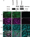
Retinal pigment epithelial (RPE) immunoreactivity is found in cultured embryonic stem (ES) cells as they differentiate. (A) Immunoblots of ES-derived RPE extract after 11 days of differentiation probed with anti-RPE65 and anti-bestrophin antibodies. Human adult RPE and fetal RPE extracts were used as positive controls. Molecular weights of RPE65 and bestrophin are indicated on the left (B–M). Immunocytochemical staining of the RPE-like cells after 14 and 28 days differentiation expressed ZO-1 at the cell margin (green, B and F) and RPE65 in the cytoplasm (purple, C and G). Nucleus is stained with DAPI (cyan, D, H, and L). Negative control at 14 days differentiation (J–M) shows only nucleus staining. Scale bars denote 25 µm.
FIGURE 4
FIGURE 4
Retinal pigment epithelium-specific protein 65 kDa (RPE65) expression in the B6 embyronic stem (ES) cell-derived RPE graft in retinal sections of albino mice at 2-week posttransplantation. Immunostaining of retinal sections of BALB/cJ (albino) mice with anti-RPE65 antibody are shown (63×). PBS injected control retina with monolayer of RPE (A); B6 ES cell-derived RPE grafts in the subretinal space of an albino Pde6gtm1/Pde6gtm1 mouse (B–D). Pde6gtm1/Pde6gtm1 mice lack photoreceptor outer segments. Note the transplanted cells overlaying host RPE cells (red arrows). Melanin granules can be seen in the B6 transplant graft (C). Notice double layer splitting after inner retina was mechanically pulled to delineate transplanted B6 ES cells (C). Red arrows mark RPE cells.
FIGURE 5
FIGURE 5
Long-term survival and distribution of embryonic stem (ES) transplants. (A) Whole-mount retina of a mouse grafted with yellow fluorescent protein (YFP)-labeled ES cell-derived retinal pigment epithelial (RPE)-like cells; YFP-positive cells around the injection site (white oval). (B) Histochemical analysis of representative sections from the whole-mount retina shown in (A) revealed an YFP-positive cell located between the OS and the RPE. IS/OS, inner segment and outer segment; ONL, outer nuclear layer; INL, inner nuclear layer.
FIGURE 6
FIGURE 6
ERGs of Rpe65rd12/Rpe65rd12 mice after subretinal transplantation with ES cell-derived retinal pigment epithelial (RPE)-like cells confirm functional rescue. (A) ERG from mice after 3 months transplantation. Eyes transplanted with ES cell-derived RPE-like cells (upper) showed higher b-wave amplitudes compared with control fellow eyes (lower). Traces represent readings from different mice. (B) b-wave enhancement in mice 1 to 7 months posttransplantation, as indicated by black solid bars. b-wave enhancement is defined as the difference in maximum ERG responses of transplanted and control fellow eyes (µV). Unpaired t tests were performed for paired differences in b-wave peaks between transplanted and control eyes. At 3 and 6 months posttransplantation, ERGs from transplanted eyes show a statistically significant rescue effect (**P = 0.001 and *P = 0.038, respectively). Although the difference was not statistically significant at 4, 5, and 7 months after transplantation, the b-wave amplitudes in the transplanted eyes were consistently higher than the control fellow eyes. The number of mice analyzed per time point is indicated. ERGs were performed on both eyes (injected and control) simultaneously. There is no statistically significant difference between injected and control eyes in the other three control groups. White bar, b-wave enhancement in PBS injected mice; light-shaded bar, b-wave enhancement in mitomycin-C treated PA6 cell transplanted mice; and dark-shaded bar, b-wave enhancement in mitomycin-C treated undifferentiated ES cell transplanted mice. ES-RPE: ES cell-derived RPE-like cells, Mit-C, mitomycin-C; ES, embryonic stem; PBS, phosphate buffered saline.

Similar articles

Cited by

References

    1. Haruta M, Sasai Y, Kawasaki H, et al. In vitro and in vivo characterization of pigment epithelial cells differentiated from primate embryonic stem cells. Invest Ophthalmol Vis Sci. 2004;45:1020. - PubMed
    1. Klassen H, Sakaguchi DS, Young MJ. Stem cells and retinal repair. Prog Retin Eye Res. 2004;23:149. - PubMed
    1. Haruta M. Embryonic stem cells: Potential source for ocular repair. Semin Ophthalmol. 2005;20:17. - PubMed
    1. Aoki H, Hara A, Nakagawa S, et al. Embryonic stem cells that differentiate into RPE cell precursors in vitro develop into RPE cell monolayers in vivo. Exp Eye Res. 2006;82:265. - PubMed
    1. Banin E, Obolensky A, Idelson M, et al. Retinal incorporation and differentiation of neural precursors derived from human embryonic stem cells. Stem Cells. 2006;24:246. - PubMed

Publication types

MeSH terms