. 1991 May 1;146(9):3227-34.

Murine tumor cells transduced with the gene for tumor necrosis factor-alpha. Evidence for paracrine immune effects of tumor necrosis factor against tumors

Affiliations

Murine tumor cells transduced with the gene for tumor necrosis factor-alpha. Evidence for paracrine immune effects of tumor necrosis factor against tumors

A L Asher et al. J Immunol. .

Abstract

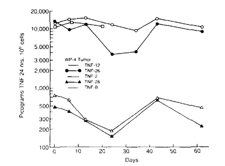
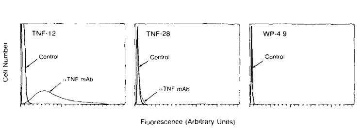
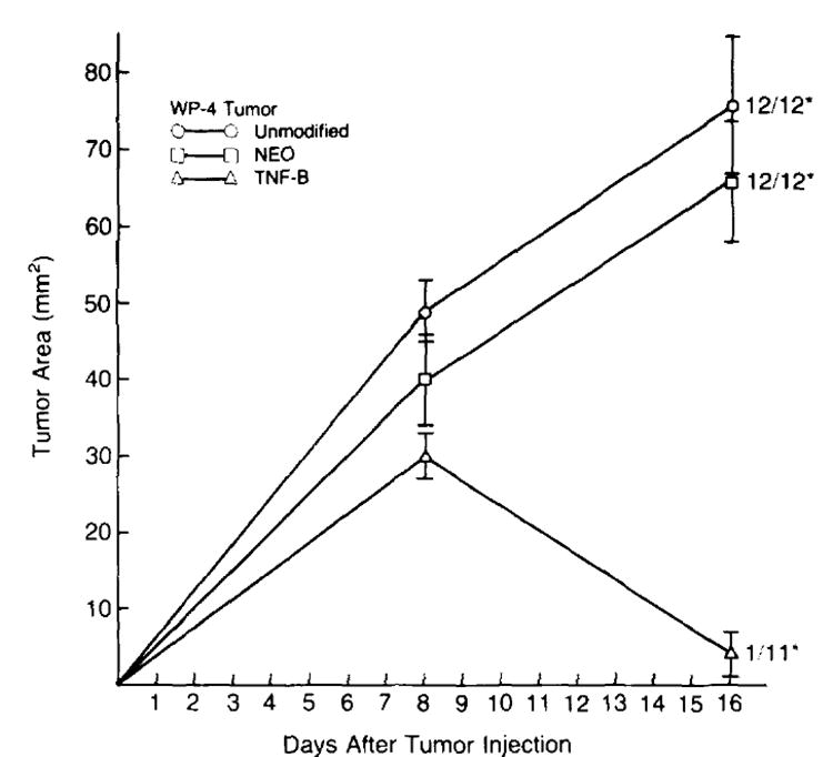
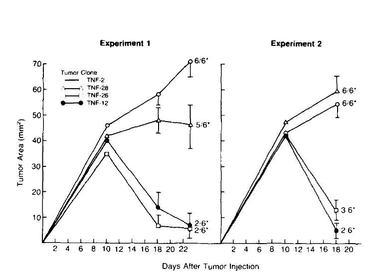
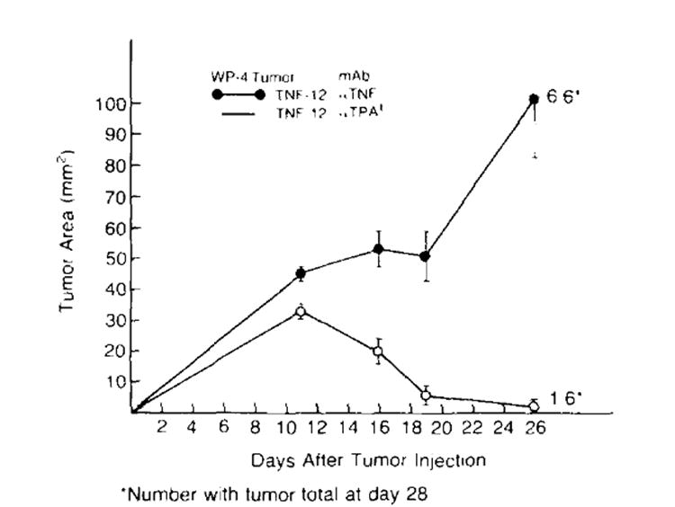
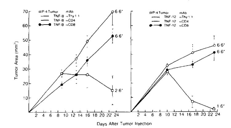
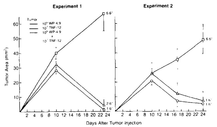
Studies of the anti-tumor activity of TNF-alpha in vivo have been hampered by the need to administer systemically toxic doses of the cytokine to obtain a curative response. To facilitate studies of the effect of high local concentrations of TNF-alpha on tumor growth and host immunity, a newly induced murine sarcoma was transduced with the gene for human TNF-alpha and the biologic characteristics of these cells were examined. We identified high and low TNF-producing tumor clones which exhibited stable TNF secretion over time. Significant amounts of membrane associated TNF were found in a high-TNF producing clone as well. No difference in the in vitro growth rates between TNF-producing and nonproducing cell lines was observed. In contrast, in vivo studies demonstrate that although unmodified parental tumor cells grew progressively when implanted s.c. in animals, tumor cells transduced with the TNF gene were found to regress in a significant number of animals after an initial phase of growth. This effect correlated with the amount of TNF produced and could be blocked with a specific anti-TNF antibody. Regressions of TNF-producing cells occurred in the absence of any demonstrable toxicity in the animals bearing these tumors. TNF-producing tumor cells could function in a paracrine fashion by inhibiting the growth of unmodified, parental tumor cells implanted at the same site. The ability of tumor cells to regress was abrogated by in vivo depletion of CD4+ or CD8+ T cell subsets and animals that had experienced regression of TNF-producing tumors rejected subsequent challenges of parental tumor. Our studies thus show that tumor cells elaborating high local concentrations of TNF regress in the absence of toxicity in the host and that this process requires the existence of intact host immunity. Studies of the lymphocytes infiltrating the gene modified tumors and attempts to use TNF gene modified tumor infiltrating lymphocytes to deliver high local concentrations of TNF to the tumor site without inducing systemic toxicity are underway.


Figures

Figure 1
Figure 1
Secretion of TNF by transduced cell lines over time. TNF production of each cell line was measured by a specific immunoassay. TNF production of various cell lines remained relatively stable over time High TNF-producing cell lines. TNF-12, TNF-26 and TNF-B (bulk transduced WP-4 tumor) made an average of 12,785 ± 843 (n = 7), 9,214 ± 1,467 (n = 7) and 11,667 ± 667 (n= 3) pg/106 cells/24 h, respectively. Low producing TNF cell lines TNF-2 and TNF-28 made a n average of 418 ± 71 (n = 6) and 455 ± 109 (n = 6) pg/106 cells/24 h, respectively. No TNF activity was detected in unmodified or neo-transfected tumor cell lines.
Figure 2
Figure 2
Flow microfluorimetric analysis of tumor cells stained with anti-human TNF antibody. A clone of the unmodified WP-4 parental culture. WP 4.9, as well as two TNF-producing clones, TNF-12 (high producer) and TNF-28 (low producer) were examined for the presence of membrane-associated human TNF. Fluorescence intensity was measured in arbitrary linear units. The 105 cells were analyzed in each frame. Negative controls stained with irrelevant antibody are represented by the sharp peaks to the far left. Cells staining positively for TNF are represented by the curves seen to the right of the controls. The high TNF producing clone was found to have significant amounts of membrane associated TNF (95% of the cells stained positively for TNF). In comparison, TNF-28, a low TNF secreter was found to express a very low amount of surface TNF. No evidence of membrane associated TNF was found on the nontransduced cell line WP 4.9.
Figure 3
Figure 3
In vivo growth of unmodified and gene modified tumor cells. B6 animals were injected s.c. with 1 × 10⁷ neo-transfected (NEO) , TNF-transduced (TNF-B).or unmodified WP-4 tumor cells on day 0 and tumor growth was followed over time. Unmodified and neo-transfected WP-4 tumors grew progressively over time and no regressions were observed. In contrast, bulk TNF-transduced tumor cells typically grew progressively over an 8- to 12-day period at which point approximately 70% regressed completely. In the representative experiment shown here, 91% of the TNF-transduced tumors regressed. (* number with tumor/total at day 28. Tumor area measurements are plotted for each time point and are expressed as mean ± SEM.)
Figure 4
Figure 4
Growth of TNF producing tumor clones in mice. Two consecutive experiments in which the in vivo growth of different TNF-producing clones was assessed. On day 0, B6 mice received 1 × 10⁷ tumor cells s.c. and tumor growth was followed over time. In both experiments, low TNF-producing clones (TNF-2 and TNF-28) grew progressively in all but one animal. In contrast, animals bearing high TNF producing tumors (TNF-12. TNF-26) experienced significant reductions in average tumor size after an initial period of tumor growth, and several animals experienced complete tumor regression. (* number with tumor/total at day 28, Tumor area measurements are plotted for each time point and are shown mean ± SEM.)
Figure 5
Figure 5
Anti-TNF antibody blocks the regression of a TNF-producing tumor. TNF-12 cells (1 × 10⁷) (a high TNF-producing clone) were inoculated s.c. into B6 mice on day 0. The animals were then divided into two experimental groups. Group one was given 100 μg of a specific anti-human TNF antibody i.p. q 3 to 4 days. Group two was treated in the same fashion with an identical quantity of control irrelevant antibody (+mouse anti-hamster tissue plasminogen activator). Tumor growth was followed. Administration of anti-TNF antibody resulted in progressive growth of TNF-producing tumors. In contrast, control antibody had no effect on the ability of these tumors to regress after an initial period of growth. (*number with tumor/total at day 28. Tumor area measurements are plotted for each time point and are shown mean ± SEM.)
Figure 6
Figure 6
In vivo depletion of CD4+ or CD8+ T cell subsets eliminates regression of TNF-producing tumors. B6 mice received a single i.v. dose of either anti-CD4. anti-CD8 or anti-Thy-1.1 mAb on day 0. Two hours after treatment with mAb, mice were inoculated s.c. with 1 × 10⁷ TNF-producing tumor cells (in experiment 1, TNF-B: in experiment 2. TNF-12) and tumor growth was followed. In both experiments, elimination of CD4+ or CD8+ cells abrogated the ability of TNF-producing cells to regress. In contrast, animals treated with anti-Thy-l.1 control mAb experienced significant reductions in tumor sizes after an initial phase of tumor growth and 9/12 animals went on to experience complete tumor regression. (*number with tumor/total at day 28. Tumor area measurements are plotted for each time point and are shown mean ± SEM.)
Figure 7
Figure 7
TNF-producing tumor cells inhibit the growth of unmodified tumor cells inoculated at the same site. The 1 × 10⁶ WP-4.9 tumor cells (a clone of the unmodified parental culture WP-4) and 1 × 10⁷ TNF-12 tumor cells (a high TNF-producing clone) were injected s.c. on day 0 into B6 animals either alone or mixed together and tumor growth was followed. Although WP-4.9 tumor cells injected alone grew progressively in all animals, animals bearing TNF-12 cells or the mixture of TNF producing and nonproducing cells experienced regression of tumor after an initial phase of tumor growth. (*number with tumor/total at day 28. Tumor measurements are plotted for each time point and are shown mean ± SEM)

References

    1. Carswell EA, Old LJ, Kassel RL, Green S, Fiore N, Williamson B. An endotoxin-induced serum factor that causes necrosis of tumors. Proc Natl Acad Sci USA. 1975;72:3666. - PMC - PubMed
    1. Asher AL, Mulé JJ, Reichert CM, Shiloni E, Rosenberg SA. Studies on the anti-tumor efficacy of systemically administered recombinant tumor necrosis factor against several murine tumors in vivo. J Immunol. 1987;138:963. - PubMed
    1. Haranaka K, Satomi N. Cytotoxic activity of tumor necrosis factor (TNF) on human cancer cells in vitro. Jpn J Exp Med. 1981;51:191. - PubMed
    1. Creasy AA, Reynolds TR, Laird W. Cures and partial regression of murine and human tumors by recombinant tumor necrosis factor. Cancer Res. 1986;46:5687. - PubMed
    1. Haranaka K, Satomi N, Sakurai A. Antitumor activity of murine tumor necrosis factor (TNF) against transplanted murine tumors and hetero-transplanted human tumors in nude mice. Int J Cancer. 1984;34:263. - PubMed

Substances

LinkOut - more resources