. 2008 Jan 1:1:1.
doi: 10.2174/1876386300801010001.

Tumor Necrosis Factor-α Suppresses Activation of Sustained Potassium Currents in Rat Small Diameter Sensory Neurons

Affiliations

Tumor Necrosis Factor-α Suppresses Activation of Sustained Potassium Currents in Rat Small Diameter Sensory Neurons

Bao-Gang Liu et al. Open Pain J. .

Abstract

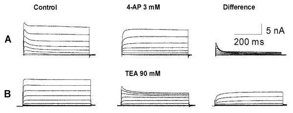
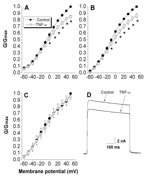
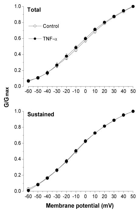
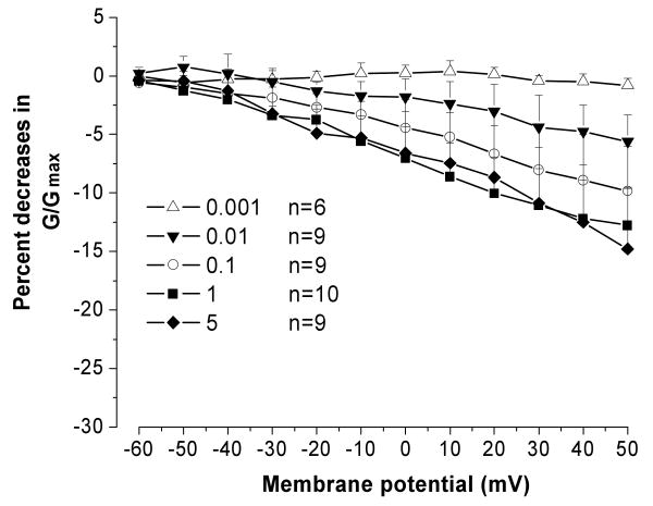
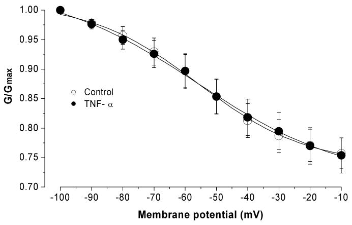
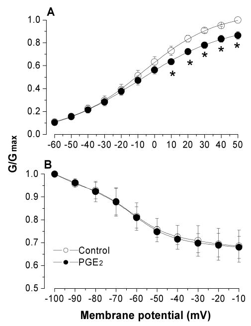
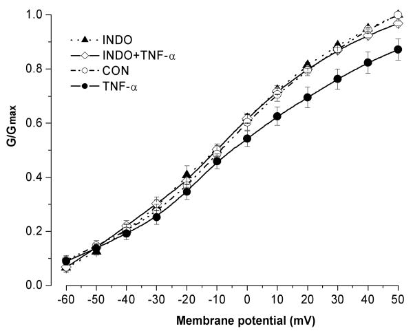
Tumor necrosis factor-α (TNF-α), a pro-inflammatory cytokine, produces pain and hyperalgesia by activating and/or sensitizing nociceptive sensory neurons. In the present study, using whole-cell patch clamp techniques, the regulation of potassium currents by TNF-α was examined in acutely dissociated small dorsal root ganglion neurons. We found that acute application of TNF-α inhibited, in a dose-dependent manner, the non-inactivating sustained potassium current without changing the rapidly inactivating transient current or the kinetics of steady-state inactivation. The effects of TNF-α on potassium currents were similar to that of prostaglandin E2 as reported previously and also demonstrated in the current study. Furthermore, indomethacin, a potent inhibitor for both cyclo-oxygenase (COX) -1 and COX-2, completely blocked the effect of TNF-α on potassium currents. These results suggest that TNF-α may sensitize or activate sensory neurons by suppressing the sustained potassium current in nociceptive DRG neurons, possibly via stimulating the synthesis/release of endogenous prostaglandins.


Figures

Figure 1
Figure 1
Pharmacological isolation of the K current reveals 2 distinct classes of small DRG neurons. A: A typical neuron with a large amount of non-inactivating sustained component and a smaller component of rapidly inactivating transient component. B: A typical small neuron with both non-inactivating sustained and fast inactivating components. Note that the rapidly inactivating component is sensitive to 4-AP; and the non-inactivating sustained component is blocked by TEA.
Figure 2
Figure 2
Effects of TNF-α on potassium currents. TNF-α (1 ng/ml) suppressed the activation of total (A) and sustained K currents (B), but did not change the rapidly inactivating transient current (C). The K current was normalized by the maximum conductance, and fitted by the Boltzmann equation, and plotted as a function of membrane potential. The activation protocol consisted of 500-ms depolarizing voltage steps to potentials between −60 and 50 mV after a 1-s voltage step to either −120 (A) or −30 mV (B). Currents were normalized by the Gmax obtained from control at +50 mV.
Figure 3
Figure 3
The potassium current suppression by TNF-α is not associated with a shift in the voltage dependence for activation. Currents from control and TNF-α treatment were independently normalized by its own Gmax obtained at +50 mV and plotted as a function of membrane potential.
Figure 4
Figure 4
Acute bath application of TNF-α did not change the steady-state inactivation of the total K current. Current was normalized by the maximum conductance and fitted by the Boltzmann equation, and plotted as a function of membrane potential. The inactivation protocol consisted of a 1-s voltage step to potentials from -100 to -10 mV followed by depolarizing voltage step to +50 mV.
Figure 5
Figure 5
The dose-dependence of the inhibitory effect of TNF-α on non-inactivating sustained K current. TNF-α-induced K current suppression was tested at different concentrations (0.001-5 ng/ml). Each data point represents the percent decrease of the normalized current (G/Gmax) over control after TNF-α application.
Figure 6
Figure 6
Acute application of PGE2 for 5 minute inhibited the non-inactivating K current without affecting steady-state inactivation.
Figure 7
Figure 7
Pretreatment of the DRG cells with indomethacin completely blocked TNF-α-induced K current inhibition. Indomethacin alone had no effect on the potassium currents.

Similar articles

Cited by

References

    1. Ahn SH, Cho YW, Ahn MW, Jang SH, Sohn YK, Kim HS. mRNA expression of cytokines and chemokines in herniated lumbar intervertebral discs. Spine. 2002;27:911–917. - PubMed
    1. Amir R, Devor M. Spike-evoked suppression and burst patterning in dorsal root ganglion neurons of the rat. J Physiol (Lond) 1997;501:183–196. - PMC - PubMed
    1. Budde T, Mager R, Pape HC. Different Types of Potassium Outward Current in Relay Neurons Acutely Isolated from the Rat Lateral Geniculate Nucleus. Eur J Neurosci. 1992;4:708–722. - PubMed
    1. Chen DB, Yang ZM, Hilsenrath R, Le SP, Harper MJ. Stimulation of prostaglandin (PG) F2 alpha and PGE2 release by tumour necrosis factor-alpha and interleukin-1 alpha in cultured human luteal phase endometrial cells. Hum Reprod. 1995;10:2773–2780. - PubMed
    1. Creange A, Barlovatz-Meimon G, Gherardi RK. Cytokines and peripheral nerve disorders. Eur Cytokine Netw. 1997;8:145–151. - PubMed

LinkOut - more resources