

Membrane Electromechanics in Biology, with a Focus on Hearing
- PMID: 20165559
- PMCID: PMC2822359
- DOI: 10.1557/mrs2009.178
Membrane Electromechanics in Biology, with a Focus on Hearing
Abstract
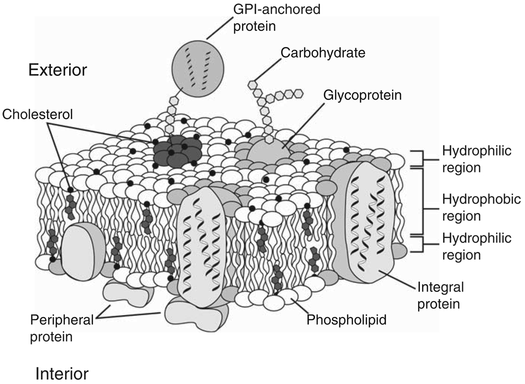
Cells are ion conductive gels surrounded by a ~5-nm-thick insulating membrane, and molecular ionic pumps in the membrane establish an internal potential of approximately -90 mV. This electrical energy store is used for high-speed communication in nerve and muscle and other cells. Nature also has used this electric field for high-speed motor activity, most notably in the ear, where transduction and detection can function as high as 120 kHz. In the ear, there are two sets of sensory cells: the "inner hair cells" that generate an electrical output to the nervous system and the more numerous "outer hair cells" that use electromotility to counteract viscosity and thus sharpen resonance to improve frequency resolution. Nature, in a remarkable exhibition of nanomechanics, has made out of soft, aqueous materials a microphone and high-speed decoder capable of functioning at 120 kHz, limited only by thermal noise. Both physics and biology are only now becoming aware of the material properties of biomembranes and their ability to perform work and sense the environment. We anticipate new examples of this biopiezoelectricity will be forthcoming.
Figures
Similar articles
-
Investigating outer hair cell motility with a combination of external alternating electrical field stimulation and high-speed image analysis.J Vis Exp. 2011 Jul 18;(53):2965. doi: 10.3791/2965. J Vis Exp. 2011. PMID: 21788937 Free PMC article.
-
Analysis of outer hair cell electromechanics reveals power delivery at the upper-frequency limits of hearing.J R Soc Interface. 2022 Jun;19(191):20220139. doi: 10.1098/rsif.2022.0139. Epub 2022 Jun 8. J R Soc Interface. 2022. PMID: 35673856 Free PMC article.
-
Outer hair cell electromechanics as a problem in soft matter physics: Prestin, the membrane and the cytoskeleton.Hear Res. 2022 Sep 15;423:108426. doi: 10.1016/j.heares.2021.108426. Epub 2021 Dec 31. Hear Res. 2022. PMID: 35101286 Review.
-
The physics of hearing: fluid mechanics and the active process of the inner ear.Rep Prog Phys. 2014 Jul;77(7):076601. doi: 10.1088/0034-4885/77/7/076601. Epub 2014 Jul 9. Rep Prog Phys. 2014. PMID: 25006839 Review.
-
Megahertz Sampling of Prestin (SLC26a5) Voltage-Sensor Charge Movements in Outer Hair Cell Membranes Reveals Ultrasonic Activity that May Support Electromotility and Cochlear Amplification.J Neurosci. 2023 Apr 5;43(14):2460-2468. doi: 10.1523/JNEUROSCI.2033-22.2023. Epub 2023 Mar 3. J Neurosci. 2023. PMID: 36868859 Free PMC article.
Cited by
-
Adaptation Independent Modulation of Auditory Hair Cell Mechanotransduction Channel Open Probability Implicates a Role for the Lipid Bilayer.J Neurosci. 2016 Mar 9;36(10):2945-56. doi: 10.1523/JNEUROSCI.3011-15.2016. J Neurosci. 2016. PMID: 26961949 Free PMC article.
-
The remarkable cochlear amplifier.Hear Res. 2010 Jul;266(1-2):1-17. doi: 10.1016/j.heares.2010.05.001. Hear Res. 2010. PMID: 20541061 Free PMC article.
-
On the Coupling between Mechanical Properties and Electrostatics in Biological Membranes.Membranes (Basel). 2021 Jun 28;11(7):478. doi: 10.3390/membranes11070478. Membranes (Basel). 2021. PMID: 34203412 Free PMC article. Review.
-
WITHDRAWN: Membrane-based amplification in hearing.Hear Res. 2009 Oct 7:10.1016/j.heares.2009.09.016. doi: 10.1016/j.heares.2009.09.016. Online ahead of print. Hear Res. 2009. PMID: 19818390 Free PMC article.
-
Electromechanical and elastic probing of bacteria in a cell culture medium.Nanotechnology. 2012 Jun 22;23(24):245705. doi: 10.1088/0957-4484/23/24/245705. Epub 2012 May 28. Nanotechnology. 2012. PMID: 22641388 Free PMC article.
References
Grants and funding
LinkOut - more resources
Full Text Sources