

Extramedullary hemopoiesis with undiagnosed, early myelofibrosis causing spastic compressive myelopathy: Case report and review
- PMID: 20165685
- PMCID: PMC2822429
- DOI: 10.4103/0019-5413.57281
Extramedullary hemopoiesis with undiagnosed, early myelofibrosis causing spastic compressive myelopathy: Case report and review
Abstract
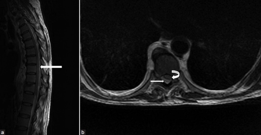
Extramedullary hemopoiesis (EMH) is a common compensatory phenomenon associated with chronic hemolytic anemia. Abnormal hemopoietic tissue usually develops in sites responsible for fetal hemopoiesis, such as spleen, liver and kidney; however, other regions such as the spine may also become involved. In this study, a patient presenting with spastic paraparesis due to EMH in the dorsal spine is described. A 62-year-old man presented with paraparesis. Magnetic resonance imaging revealed a large lesion involving the T2-L2 vertebral levels with a large extradural component causing thecal sac compression. Laminectomy with excision of mass was carried out. The histopathology revealed EMH. The patient had no known cause for EMH at the time of diagnosis but, subsequently, a bone marrow examination revealed early myelofibrosis. This case represents the rare occurrence of a large extradural extramedullary hematopoiesis in a patient with no known predisposing factor for hemopoiesis at the time of presentation.
Keywords: Extramedullary hemopoiesis; myelofibrosis; spastic paraparesis; spine.
Conflict of interest statement
Figures

Similar articles
-
[Spinal cord compression due to extramedullary hematopoiesis in a patient with myelofibrosis].Rinsho Shinkeigaku. 2014;54(1):27-31. doi: 10.5692/clinicalneurol.54.27. Rinsho Shinkeigaku. 2014. PMID: 24429645 Japanese.
-
Huge extramedullary hematopoiesis mass in the posterior mediastinum: a case report.Front Oncol. 2024 Dec 13;14:1489785. doi: 10.3389/fonc.2024.1489785. eCollection 2024. Front Oncol. 2024. PMID: 39735597 Free PMC article.
-
Compressive Dorsal Myelopathy Secondary to Extramedullary Hematopoiesis in a Thalassemic Patient.Case Rep Neurol Med. 2019 Oct 17;2019:5827626. doi: 10.1155/2019/5827626. eCollection 2019. Case Rep Neurol Med. 2019. PMID: 31781438 Free PMC article.
-
Extramedullary hemopoiesis.Semin Ultrasound CT MR. 2014 Jun;35(3):255-62. doi: 10.1053/j.sult.2013.12.001. Epub 2013 Dec 19. Semin Ultrasound CT MR. 2014. PMID: 24929265
-
Cutaneous extramedullary hemopoiesis in chronic myeloproliferative and myelodysplastic disorders.J Am Acad Dermatol. 2006 Aug;55(2 Suppl):S28-31. doi: 10.1016/j.jaad.2005.11.1038. J Am Acad Dermatol. 2006. PMID: 16843120 Review.
References
-
- Tai SM, Chan JS, Ha SY, Young BW, Chan MS. Successful treatment of spinal cord compression secondary to extramedullary haemopoietic mass by hypertransfusion in a patient with thalassemia major. Pediatr Hematol Oncol. 2006;23:317–21. - PubMed
-
- Niggeman P, Krings T, Hans F, Thron A. Fifteen-year follow-up of a patient with beta thalassaemia and extramedullary haematopoietic tissue compressing the spinal cord. Neuroradiology. 2005;47:263–6. - PubMed
-
- Salehi SA, Koski T, Ondra SL. Spinal cord compression in beta-thalassemia: Case report and review of the literature. Spinal Cord. 2004;42:117–23. - PubMed
-
- Hiradfur M, Zabolinejadm N, Banihashem A, Kajbafzadeh AM. Renal splenic heterotopia with extramedullary haemopoiesis in a thalassemic patient, simulating renal neoplasm: A case report. J Pediatr Hematol Oncol. 2007;29:195–7. - PubMed
-
- Sauer B, Buy X, Gangi X, Roy C. Exceptional localization of extramedullary haemopoiesis: Presacral and periureteral masses. Acta Radiol. 2007;48:246–8. - PubMed
LinkOut - more resources
Full Text Sources