

Primary synovial osteochondromatosis of a subdeltoid bursa
- PMID: 20165686
- PMCID: PMC2822411
- DOI: 10.4103/0019-5413.58613
Primary synovial osteochondromatosis of a subdeltoid bursa
Abstract
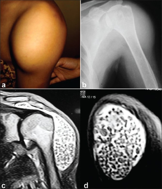
Primary synovial osteochondromatosis (SOC) is known to be intra-articular and wherever it is observed outside a synovial joint, it is associated with the involvement of the nearby joint. Primary SOC has not been reported to involve a subdeltoid bursa. We present a case of a 52-year-old woman having a large number of loose bodies in a large tumor in the subdeltoid bursa. The swelling was first noticed by the patient 2 years back. Plain roentgenogram revealed soft tissue swelling only with no areas of calcification. On MRI, multiple nonosseous loose bodies were visualized in the bursa deep to the deltoid muscle. A surgical excision of subdeltoid bursa was done. A biopsy confirmed it to be cartilaginous loose bodies in synovial lining sugestive of metaplastic transformation of the synovial tissue.
Keywords: Subdeltoid bursa; extraarticular synovial osteochondromatosis; synovial osteochondromatosis.
Conflict of interest statement
Figures


References
-
- Henderson MS, Joxis HT. Loose bodies in joints and bursae due to synovial osteochondromatosis. J Bone Joint Surg. 1923;5:400–4.
-
- Jones HT. Loose body formation in synovial osteochondromatosis with special reference to the etiology and pathology. J Bone Joint Surg. 1924;6:407–58.
-
- Mussey RD, Henderson MS. Osteochondromatosis. J Bone Joint Surg Am. 1949;31:619–27. - PubMed
-
- Jeffreys TE. Synovial chondromatosis. J Bone Joint Surg Br. 1967;49:530–4. - PubMed
-
- Dorfman HD. In “Bone Tumors”. Mosby; 1998. Czerniak: Synovial chondromatosis. In synovial lesions; pp. 1041–57.
LinkOut - more resources
Full Text Sources