Review
. 2010 Jun;67(12):2001-24.
doi: 10.1007/s00018-010-0299-5. Epub 2010 Feb 18.

In front of and behind the replication fork: bacterial type IIA topoisomerases

Affiliations
Review

In front of and behind the replication fork: bacterial type IIA topoisomerases

Claudia Sissi et al. Cell Mol Life Sci. 2010 Jun.

Abstract

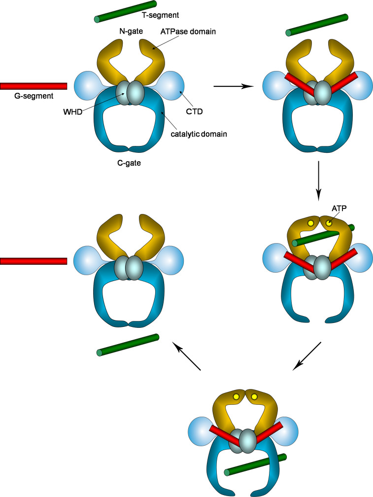
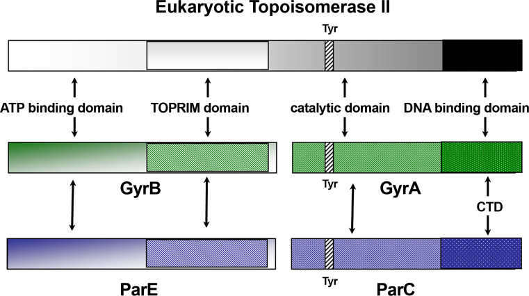
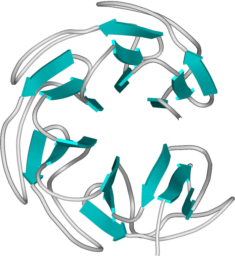
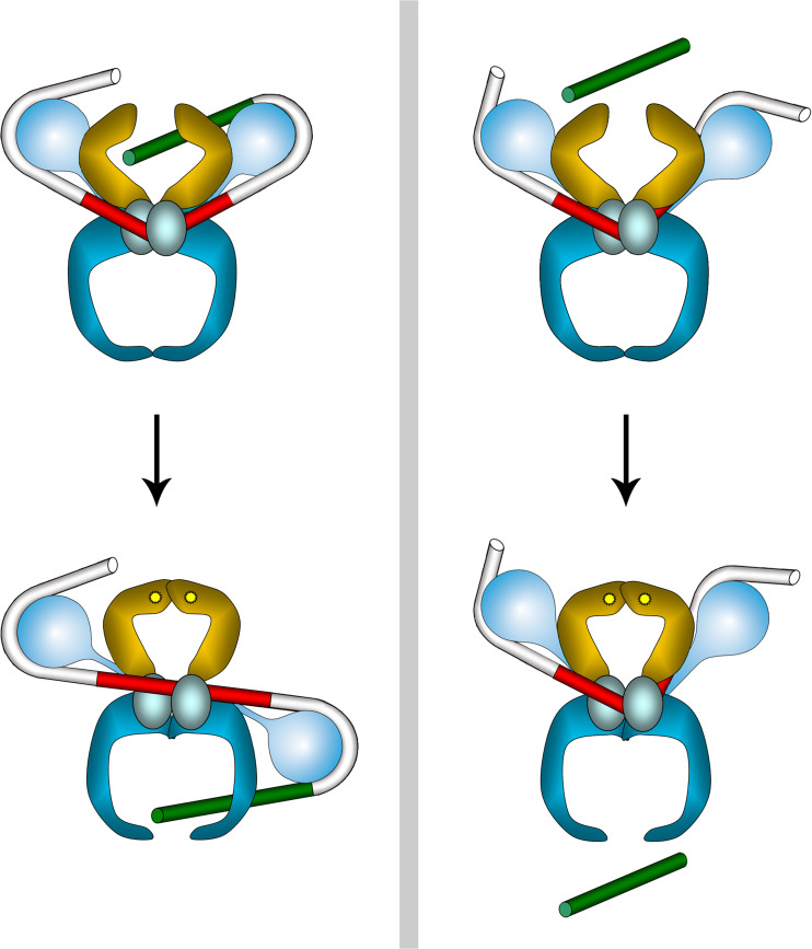
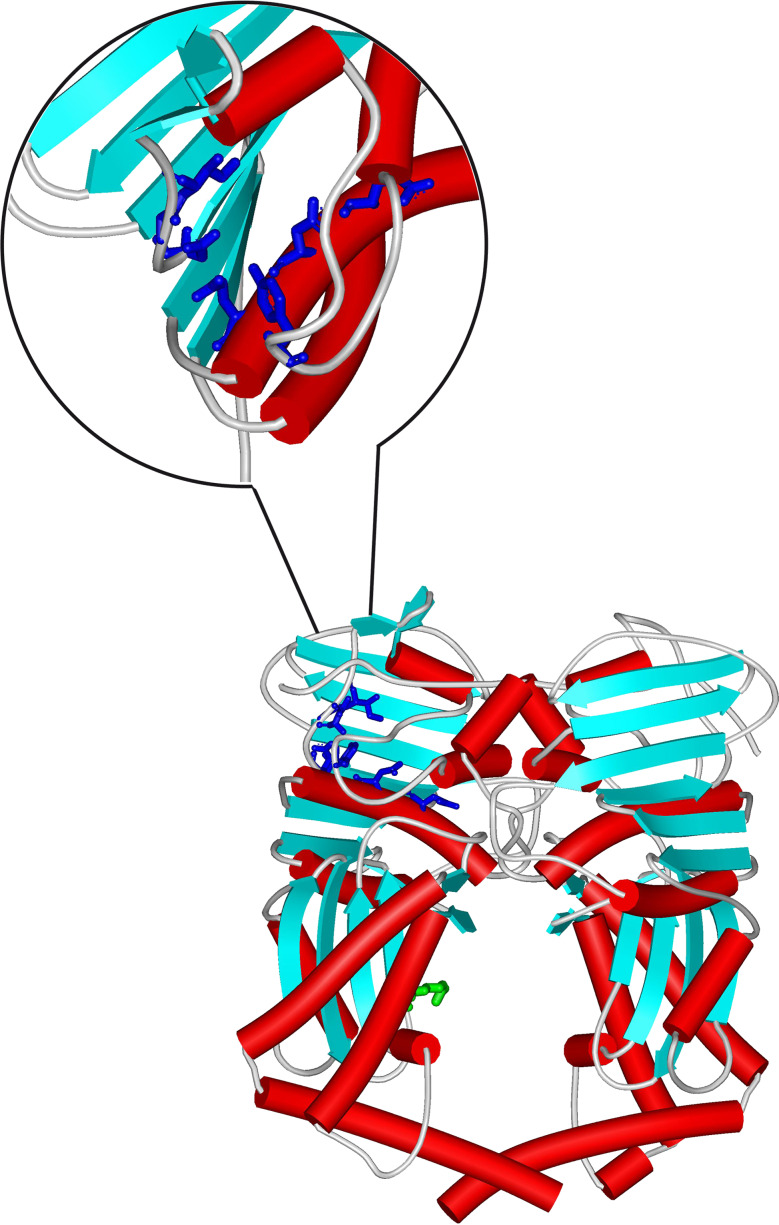
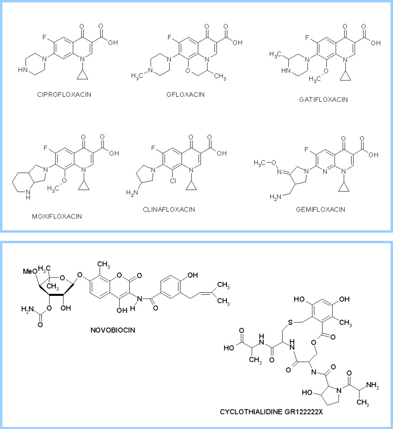
Topoisomerases are vital enzymes specialized in controlling DNA topology, in particular supercoiling and decatenation, to properly handle nucleic acid packing and cell dynamics. The type IIA enzymes act by cleaving both strands of a double helix and having another strand from the same or another molecule cross the DNA gate before a re-sealing event completes the catalytic cycle. Here, we will consider the two types of IIA prokaryotic topoisomerases, DNA Gyrase and Topoisomerase IV, as crucial regulators of bacterial cell cycle progression. Their synergistic action allows control of chromosome packing and grants occurrence of functional transcription and replication processes. In addition to displaying a fascinating molecular mechanism of action, which transduces chemical energy into mechanical energy by means of large conformational changes, these enzymes represent attractive pharmacological targets for antibacterial chemotherapy.


Figures

Fig. 1
Fig. 1
Different topological states of covalently closed circular DNA
Fig. 2
Fig. 2
Relevant steps of the two-gate mechanism for type IIA topoisomerases. CTD C-terminal domain, WHD winged-helix domain, containing the catalytic Tyr residue. The G-segment is engaged at the GyrA/GyrB or ParC/ParE domain interface, where it is sharply bent, T-segment is recruited in the ATP-clamp (N-gate), which is closed when ATP binds. Then, the G-gate opens, forming the cleavage complex and allows T-segment transport. Finally, T-segment exits through the C-gate and the enzyme is reset for a subsequent catalytic cycle
Fig. 3
Fig. 3
Schematic organization of functional domains in type IIA topoisomerases. Location of catalytic tyrosine (Tyr) is indicated
Fig. 4
Fig. 4
Crystal structure of E. coli GyrA59 dimer (PDB ID 1AB4). Red tubes refer to α-helix regions, blue arrows to β-sheet. The catalytic Tyr is highlighted in green (ball-and-stick model)
Fig. 5
Fig. 5
Crystal structure of the S. pneumoniae ParC breakage-reunion domain (ParC55) and ParE TOPRIM domain (ParE30) in the cleavage complex with DNA in the presence of the quinolone moxifloxacin (PDB ID 3FOF). Red tubes refer to α-helix regions, blue arrows to β-sheet. The DNA portion is in green sticks
Fig. 6
Fig. 6
Relative location of the WHD (cyan), tower (green), and TOPRIM (brown) domains in the catalytic DNA cleavage-resealing pocket as observed in the crystal structure of the S. pneumoniae ParC breakage-reunion domain (ParC55) in combination with the ParE TOPRIM domain (ParE30) in the cleavage complex with DNA(PDB ID 3FOF). The catalytic Tyr residue in WHD and the four acidic residues involved in Mg2+ binding in TOPRIM are highlighted (ball-and-stick model). Tubes represent α-helix regions, arrows β-sheets
Fig. 7
Fig. 7
The β-pinwheel structure of the E. coli GyrA CTD (PDB ID 1ZI0). Blue arrows refer to β- sheet tracts
Fig. 8
Fig. 8
Proposed model for diverse recruitment of G (red) and T (green) segments by DNA Gyrase (left panel) and Topoisomerase IV (right panel). Labeling as in Fig. 2
Fig. 9
Fig. 9
Crystal structure of the 43-kDa E. coli GyrB dimer in complex with ADPNP, a nonhydrolyzable analogue of ATP (PDB ID 1E11). Red tubes refer to α-helix regions, blue arrows to β-sheet. Residues Glu42, Asn46, Glu50, Asp73, Arg76, Gly77, Ile78 and Tyr109 in the ATP hydrolysis region are highlighted in dark blue (ball-and-stick models, see also close-up), Asp286 in the DNA binding region in green (see text for details)
Fig. 10
Fig. 10
Representative bacterial topoisomerase II inhibitors. Chemical structure of cleavage complex poisons (upper panel) and ATPase inhibitors (lower panel)
Fig. 11
Fig. 11
Close-up view of the quinolone moxifloxacin (stick model) bound to the S. pneumoniae ParC55-ParE30-DNA cleavage complex (line model). Source: PDB ID 3FOF

References

    1. Kalkbrenner T, Arnold A, Tans SJ. Internal dynamics of supercoiled DNA molecules. Biophys J. 2009;96:4951–4955. doi: 10.1016/j.bpj.2009.03.056. - DOI - PMC - PubMed
    1. Liu Z, Deibler RW, Chan HS, Zechiedrich L. The why and how of DNA unlinking. Nucleic Acids Res. 2009;37:661–671. doi: 10.1093/nar/gkp041. - DOI - PMC - PubMed
    1. Wasserman SA, Cozzarelli NR. Biochemical topology: applications to DNA recombination and replication. Science. 1986;232:951–960. doi: 10.1126/science.3010458. - DOI - PubMed
    1. Drolet M. Growth inhibition mediated by excess negative supercoiling: the interplay between transcription elongation, R-loop formation and DNA topology. Mol Microbiol. 2006;59:723–730. doi: 10.1111/j.1365-2958.2005.05006.x. - DOI - PubMed
    1. Ohniwa RL, Morikawa K, Kim J, Ohta T, Ishihama A, Wada C, Takeyasu K. Dynamic state of DNA topology is essential for genome condensation in bacteria. EMBO J. 2006;25:5591–5602. doi: 10.1038/sj.emboj.7601414. - DOI - PMC - PubMed

Publication types

LinkOut - more resources