Review
. 2010;17(14):1468-86.
doi: 10.2174/092986710790980005.

Endocannabinoid binding to the cannabinoid receptors: what is known and what remains unknown

Affiliations
Review

Endocannabinoid binding to the cannabinoid receptors: what is known and what remains unknown

Patricia H Reggio. Curr Med Chem. 2010.

Abstract

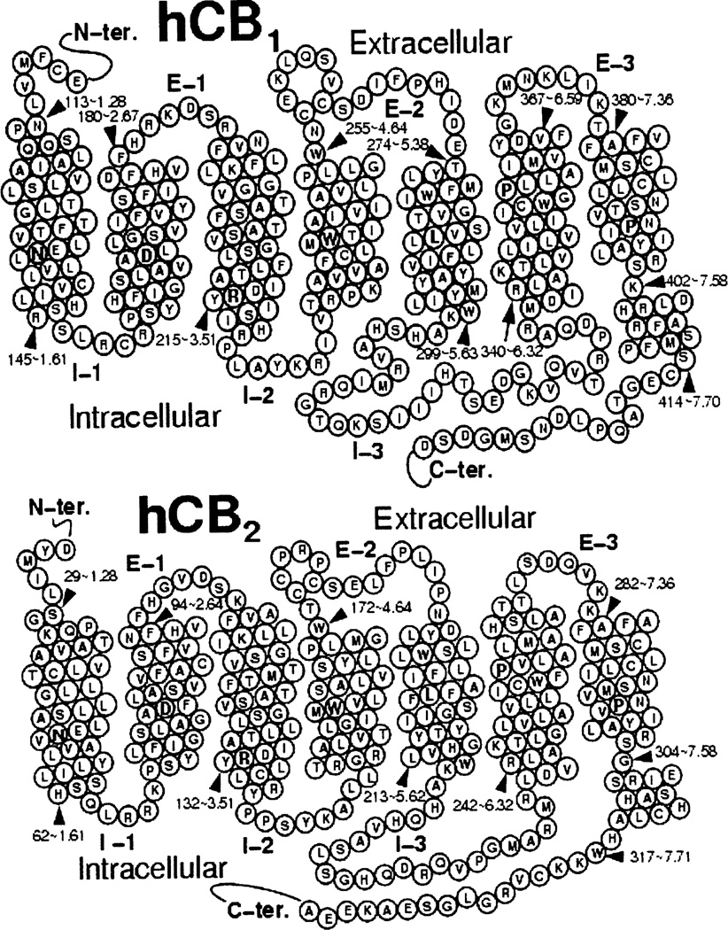
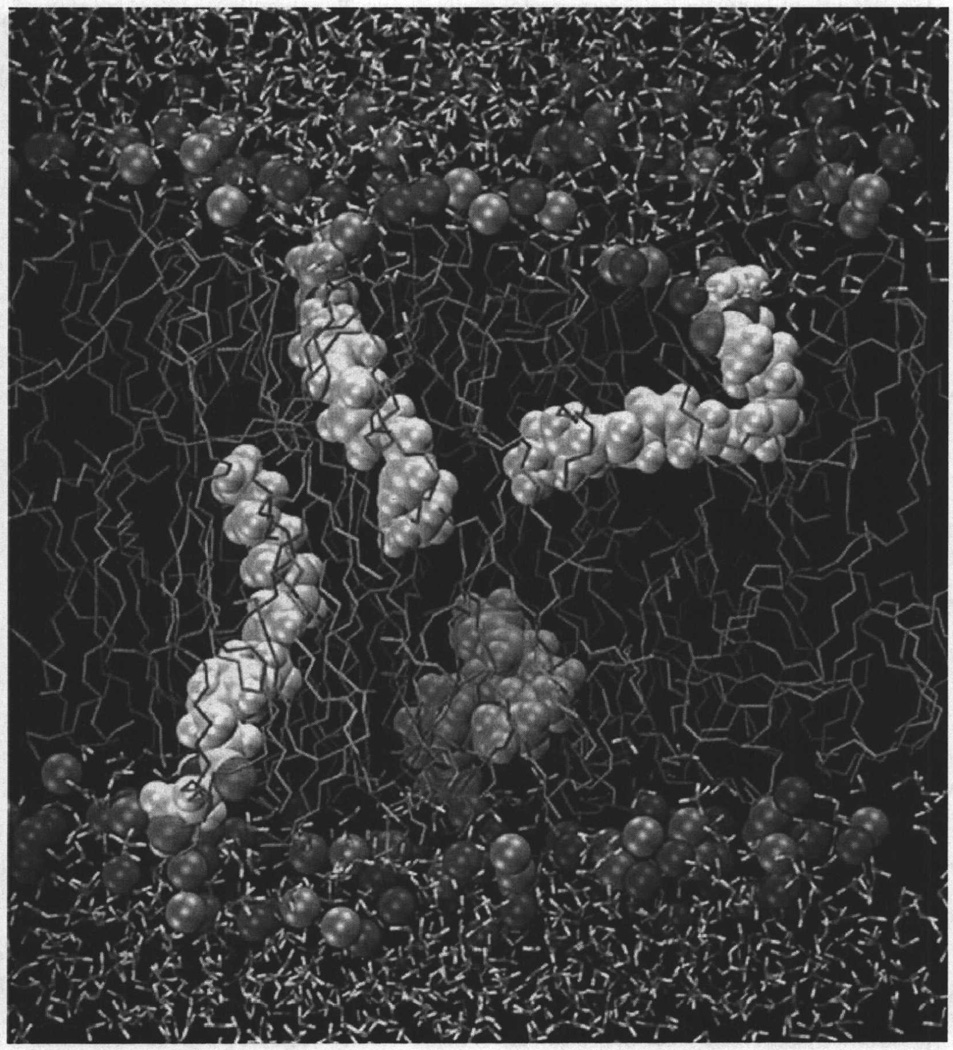
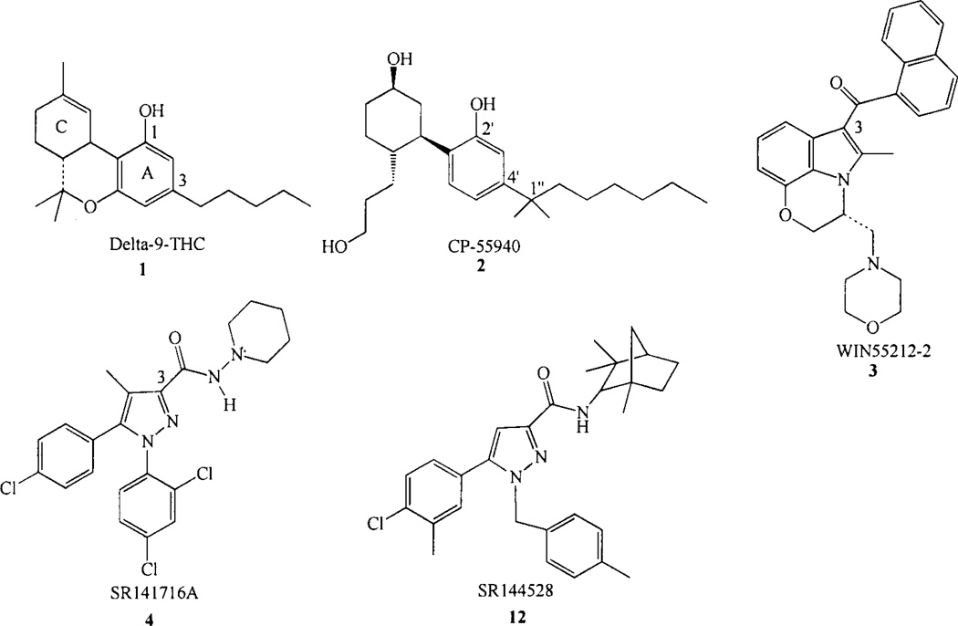
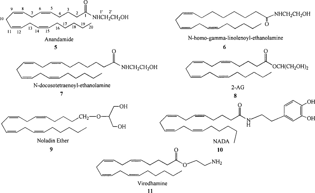
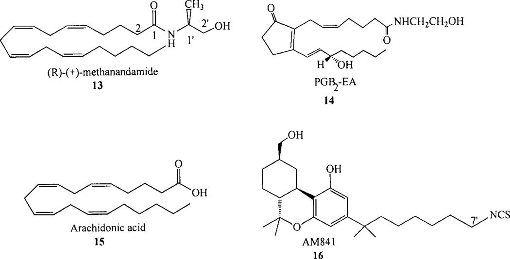
The cannabinoid CB1 and CB2 receptors are Class A G protein-coupled receptors (GPCRs). While many Class A GPCRs have endogenous ligands that are hydrophilic cations (e.g., the serotonin and dopamine receptors), the cannabinoid receptors have neutral, highly lipophilic ligands derived from the fatty acid, arachidonic acid. The most well-studied of these are N-arachidonoylethanolamine (anandamide, AEA) and sn-2-arachidonoylglycerol (2-AG). This review focuses on the experimental and computational studies that have been used to probe the nature of endocannabinoid interaction with the cannabinoid receptors. These studies include mutation, SAR and NMR studies, as well as, QSAR, docking and molecular dynamics simulations. Gaps in our knowledge are identified. The review begins more generally, however, by discussing the entire endocannabinoid system, of which the cannabinoid receptors are part. For in order to understand endocannabinoid action, one needs an appreciation for the environments for which these ligands have been designed and the conformational changes these ligands must undergo in order to act on the cannabinoid receptors.


Figures

Fig. (1)
Fig. (1)
Helix net representations of the amino acid sequences of the CB1 and CB2 receptors are presented here.
Fig. (2)
Fig. (2)
A simulation cell containing four molecules of N-arachidonoylethanolamine (anandamide, AEA, 5, Chart 2), in a fully hydrated 1,2-dioleoyl-sn-glycero-3-phosphocholine (DOPC) bilayer is illustrated here. Spheres represent the DOPC choline group nitrogen and phosphate group phosphorous atoms. The DOPC acyl chains are shown in tube display. Molecules of 5 are contoured at their Van der Waals radii, while water is depicted here in tube display. This figure shows that when 5 adopts an extended conformation in the bilayer, its terminal methyl group is at the center of the bilayer. This figure also shows that 5 is capable of forming more compact shapes while in the bilayer [210].
Fig. (3)
Fig. (3)
The AEA/R* complex in the TMH2-3-6-7 region of CB1 R* is illustrated here. AEA (5) is shown in tube display. K3.28 forms a hydrogen bond with the amide oxygen of 5. At the same time, the head group hydroxyl of 5 is engaged in an intramolecular hydrogen bond with its amide oxygen. The AEA binding pocket is lined with residues that are largely hydrophobic, including L3.29, V3.32, F6.60, F7.35, A7.36, Y6.57, S7.39 (hydrogen bonded back to its own backbone carbonyl oxygen) and L7.43. F3.25 has a C-H•••π interaction with the C5-C6 double bond of 5; while, F2.57 has an interaction with the amide oxygen of 5 [169].
Fig. (4)
Fig. (4)
A simulation cell containing a model of the CB2 receptor [211] immersed in a fully hydrated palmitoyl-oleoyl-phosphatidylcholine (POPC) lipid bilayer is illustrated here from a microsecond timescale NAMD2 MD simulation [209], The spheres represent the phosphorous atoms of the POPC head-groups. Water and the fatty acid acyl chains of the bilayer have been turned off for simplicity. 2-AG is contoured at its Van der Waals’ radius. In this figure, a 2-AG molecule has entered the CB2 receptor by passing between TMH6 and TMH7. Inset. Subsequent to entry, 2-AG establishes a long lasting interaction with D(275) in the EC-3 loop, a hydrogen bond with S6.58(268), and maintains an intramolecular hydrogen bond.
Chart 1
Chart 1
Cannabinoid receptor agonists and antagonists.
Chart 2
Chart 2
Endogenous cannabinoids.
Chart 3
Chart 3

Similar articles

Cited by

References

    1. Matsuda LA, Lolait SJ, Brownstein MJ, Young AC, Bonner TI. Structure of a cannabinoid receptor and functional expression of the cloned cDNA. Nature. 1990;346:561–564. - PubMed
    1. Felder CC, Joyce KE, Briley EM, Mansouri J, Mackie K, Blond O, Lai Y, Ma AL, Mitchell RL. Comparison of the pharmacology and signal transduction of the human cannabinoid CB1 and CB2 receptors. Mol. Pharmacol. 1995;48:443–450. - PubMed
    1. Mackie K, Lai Y, Westenbroek R, Mitchell R. Cannabinoids activate an inwardly rectifying potassium conductance and inhibit Q-type calcium currents in AtT20 cells transfected with rat brain cannabinoid receptor. J. Neurosci. 1995;15:6552–6561. - PMC - PubMed
    1. Pan X, Ikeda SR, Lewis DL. Rat brain cannabinoid receptor modulates N-type Ca2+ channels in a neuronal expression system. Mol Pharmacol. 1996;49:707–714. - PubMed
    1. Glass M, Dragunow M, Faull RL. Cannabinoid receptors in the human brain: a detailed anatomical and quantitative autoradiographic study in the fetal, neonatal and adult human brain. Neuroscience. 1997;77:299–318. - PubMed

Publication types

MeSH terms