

Keratin 19 marks poor differentiation and a more aggressive behaviour in canine and human hepatocellular tumours
- PMID: 20167095
- PMCID: PMC2834617
- DOI: 10.1186/1476-5926-9-4
Keratin 19 marks poor differentiation and a more aggressive behaviour in canine and human hepatocellular tumours
Abstract
Background: The expression of Keratin 19 (K19) was reported in a subset of hepatocellular carcinomas (HCCs). K19 positive HCCs are associated with an increased malignancy compared to K19 negative HCCs. No suitable mouse models exist for this subtype of HCC, nor is the incidence of K19 expression in hepatocellular neoplasia in model animals known. Therefore, we compared the occurrence and tumour behaviour of K19 positive hepatocellular neoplasias in dog and man.
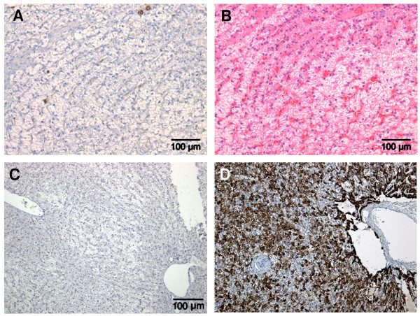
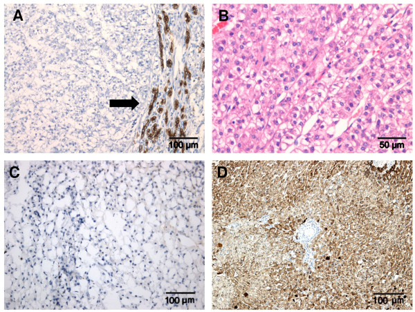
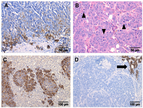
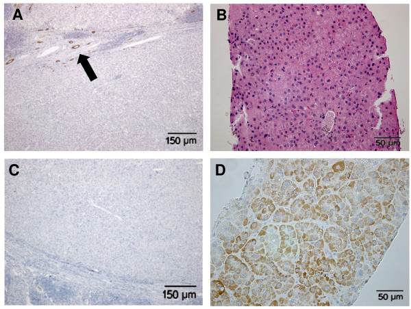
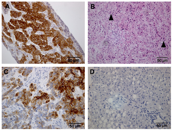
Results: The expression of hepatocellular differentiation (HepPar-1), biliary/progenitor cell (K7, K19), and malignancy (glypican-3) markers was semi-quantitatively assessed by immunohistochemistry. The histological grade of tumour differentiation was determined according to a modified classification of Edmondson and Steiner; the staging included intrahepatic, lymph node or distant metastases. Four of the 34 canine hepatocellular neoplasias showed K19 positivity (12%), of which two co-expressed K7. K19 positive tumours did not express HepPar-1, despite the histological evidence of a hepatocellular origin. Like in human HCC, all K19 positive hepatocellular neoplasias were glypican-3 positive and histologically poorly differentiated and revealed intra- or extrahepatic metastases whereas K19 negative hepatocellular neoplasias did not.
Conclusions: K19 positive hepatocellular neoplasias are highly comparable to man and occur in 12% of canine hepatocellular tumours and are associated with a poorly differentiated histology and aggressive tumour behaviour.
Figures
Similar articles
-
Cellular characteristics of keratin 19-positive canine hepatocellular tumours explain its aggressive behaviour.Vet Rec Open. 2017 Oct 21;4(1):e000212. doi: 10.1136/vetreco-2016-000212. eCollection 2017. Vet Rec Open. 2017. PMID: 29118993 Free PMC article.
-
Expression of multidrug resistance-associated protein 1 in hepatocellular carcinoma is associated with a more aggressive tumour phenotype and may reflect a progenitor cell origin.Liver Int. 2008 Dec;28(10):1370-80. doi: 10.1111/j.1478-3231.2008.01889.x. Liver Int. 2008. PMID: 19055643
-
Heterogeneity of Epigenetic and Epithelial Mesenchymal Transition Marks in Hepatocellular Carcinoma with Keratin 19 Proficiency.Liver Cancer. 2019 Jul;8(4):239-254. doi: 10.1159/000490806. Epub 2018 Aug 21. Liver Cancer. 2019. PMID: 31602368 Free PMC article.
-
Clinico-Radio-Pathological and Molecular Features of Hepatocellular Carcinomas with Keratin 19 Expression.Liver Cancer. 2020 Dec;9(6):663-681. doi: 10.1159/000510522. Epub 2020 Oct 23. Liver Cancer. 2020. PMID: 33442539 Free PMC article. Review.
-
Hepatocellular carcinomas expressing 'stemness'-related markers: clinicopathological characteristics.Dig Dis. 2014;32(6):778-85. doi: 10.1159/000368021. Epub 2014 Oct 29. Dig Dis. 2014. PMID: 25376296 Review.
Cited by
-
Cellular characteristics of keratin 19-positive canine hepatocellular tumours explain its aggressive behaviour.Vet Rec Open. 2017 Oct 21;4(1):e000212. doi: 10.1136/vetreco-2016-000212. eCollection 2017. Vet Rec Open. 2017. PMID: 29118993 Free PMC article.
-
KRT8 upregulation promotes tumor metastasis and is predictive of a poor prognosis in clear cell renal cell carcinoma.Oncotarget. 2017 Jul 12;8(44):76189-76203. doi: 10.18632/oncotarget.19198. eCollection 2017 Sep 29. Oncotarget. 2017. PMID: 29100303 Free PMC article.
-
Analysis of clinicopathologic and imaging features of dual-phenotype hepatocellular carcinoma.Sci Rep. 2024 Feb 9;14(1):3314. doi: 10.1038/s41598-024-53831-5. Sci Rep. 2024. PMID: 38332165 Free PMC article.
-
Immunotherapy for Lewis lung carcinoma utilizing dendritic cells infected with CK19 gene recombinant adenoviral vectors.Oncol Rep. 2015 Nov;34(5):2289-95. doi: 10.3892/or.2015.4231. Epub 2015 Aug 27. Oncol Rep. 2015. PMID: 26323510 Free PMC article.
-
Hepatic progenitor cells in canine and feline medicine: potential for regenerative strategies.BMC Vet Res. 2014 Jun 19;10:137. doi: 10.1186/1746-6148-10-137. BMC Vet Res. 2014. PMID: 24946932 Free PMC article. Review.
References
LinkOut - more resources
Full Text Sources
Research Materials