

Exposure of beta-tubulin regions defined by antibodies on an Arabidopsis thaliana microtubule protofilament model and in the cells
- PMID: 20167106
- PMCID: PMC2844066
- DOI: 10.1186/1471-2229-10-29
Exposure of beta-tubulin regions defined by antibodies on an Arabidopsis thaliana microtubule protofilament model and in the cells
Abstract
Background: The function of the cortical microtubules, composed of alphabeta-tubulin heterodimers, is linked to their organizational state which is subject to spatial and temporal modulation by environmental cues. The role of tubulin posttranslational modifications in these processes is largely unknown. Although antibodies against small tubulin regions represent useful tool for studying molecular configuration of microtubules, data on the exposure of tubulin epitopes on plant microtubules are still limited.
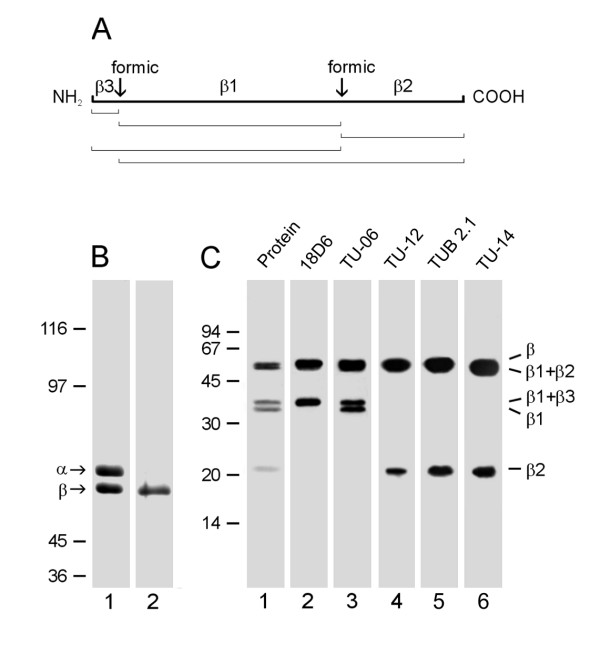
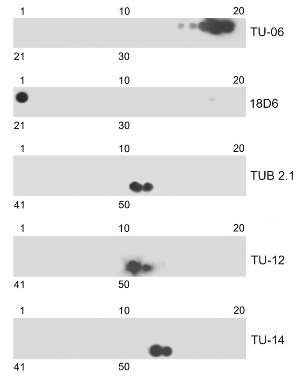
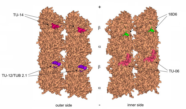
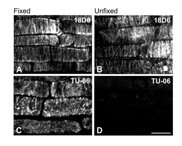
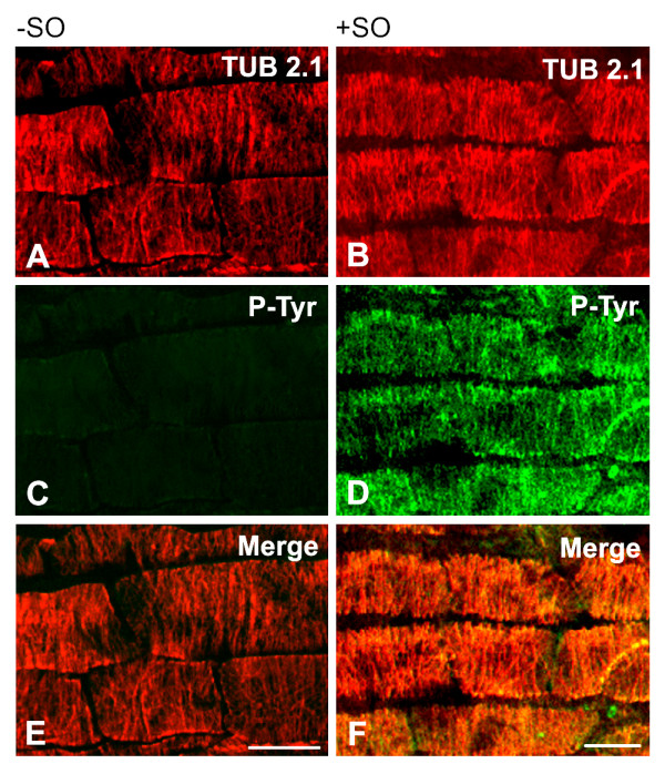
Results: Using homology modeling we have generated an Arabidopsis thaliana microtubule protofilament model that served for the prediction of surface exposure of five beta-tubulin epitopes as well as tyrosine residues. Peptide scans newly disclosed the position of epitopes detected by antibodies 18D6 (beta1-10), TUB2.1 (beta426-435) and TU-14 (beta436-445). Experimental verification of the results by immunofluorescence microscopy revealed that the exposure of epitopes depended on the mode of fixation. Moreover, homology modeling showed that only tyrosines in the C-terminal region of beta-tubulins (behind beta425) were exposed on the microtubule external side. Immunofluorescence microscopy revealed tyrosine phosphorylation of microtubules in plant cells, implying that beta-tubulins could be one of the targets for tyrosine kinases.
Conclusions: We predicted surface exposure of five beta-tubulin epitopes, as well as tyrosine residues, on the surface of A. thaliana microtubule protofilament model, and validated the obtained results by immunofluorescence microscopy on cortical microtubules in cells.The results suggest that prediction of epitope exposure on microtubules by means of homology modeling combined with site-directed antibodies can contribute to a better understanding of the interactions of plant microtubules with associated proteins.
Figures
Similar articles
-
Exposure of tubulin structural domains in Nicotiana tabacum microtubules probed by monoclonal antibodies.Eur J Cell Biol. 1997 Feb;72(2):104-12. Eur J Cell Biol. 1997. PMID: 9157006
-
Heterogeneity of microtubules recognized by monoclonal antibodies to alpha-tubulin.Eur J Cell Biol. 1986 Jun;41(1):82-8. Eur J Cell Biol. 1986. PMID: 3792338
-
Carboxy-terminal regions on the surface of tubulin and microtubules. Epitope locations of YOL1/34, DM1A and DM1B.J Mol Biol. 1986 May 20;189(2):367-70. doi: 10.1016/0022-2836(86)90517-6. J Mol Biol. 1986. PMID: 2427729
-
A microtubule bestiary: structural diversity in tubulin polymers.Mol Biol Cell. 2017 Nov 1;28(22):2924-2931. doi: 10.1091/mbc.E16-05-0271. Mol Biol Cell. 2017. PMID: 29084910 Free PMC article. Review.
-
Microtubules: an overview.Methods Mol Med. 2007;137:1-16. doi: 10.2119/molecular%20medicine-2006-00038. Methods Mol Med. 2007. PMID: 18085218 Review.
Cited by
-
Plant microtubules reorganization under the indirect UV-B exposure and during UV-B-induced programmed cell death.Plant Signal Behav. 2013 May;8(5):e24031. doi: 10.4161/psb.24031. Epub 2013 Feb 25. Plant Signal Behav. 2013. PMID: 23438586 Free PMC article.
-
Tubulin tyrosine nitration regulates microtubule organization in plant cells.Front Plant Sci. 2013 Dec 26;4:530. doi: 10.3389/fpls.2013.00530. eCollection 2013. Front Plant Sci. 2013. PMID: 24421781 Free PMC article.
-
Genome-wide identification and evolution of the tubulin gene family in Camelina sativa.BMC Genomics. 2024 Jun 14;25(1):599. doi: 10.1186/s12864-024-10503-y. BMC Genomics. 2024. PMID: 38877397 Free PMC article.
-
Members of the Plant CRK Superfamily Are Capable of Trans- and Autophosphorylation of Tyrosine Residues.J Biol Chem. 2015 Jul 3;290(27):16665-77. doi: 10.1074/jbc.M114.617274. Epub 2015 May 12. J Biol Chem. 2015. PMID: 25969537 Free PMC article.
-
Autophagy formation, microtubule disorientation, and alteration of ATG8 and tubulin gene expression under simulated microgravity in Arabidopsis thaliana.NPJ Microgravity. 2024 Mar 18;10(1):31. doi: 10.1038/s41526-024-00381-9. NPJ Microgravity. 2024. PMID: 38499552 Free PMC article.
References
Publication types
MeSH terms
Substances
LinkOut - more resources
Full Text Sources
Molecular Biology Databases
