. 2010 Feb 18:7:12.
doi: 10.1186/1476-9255-7-12.

Role of PPAR-delta in the development of zymosan-induced multiple organ failure: an experiment mice study

Affiliations

Role of PPAR-delta in the development of zymosan-induced multiple organ failure: an experiment mice study

Maria Galuppo et al. J Inflamm (Lond). .

Abstract

Background: Peroxisome proliferator-activated receptor (PPAR)-beta/delta is a nuclear receptor transcription factor that regulates gene expression in many important biological processes. It is expressed ubiquitously, especially white adipose tissue, heart, muscle, intestine, placenta and macrophages but many of its functions are unknown. Saturated and polyunsaturated fatty acids activate PPAR-beta/delta, but physiological ligands have not yet been identified. In the present study, we investigated the anti-inflammatory effects of PPAR-beta/delta activation, through the use of GW0742 (0,3 mg/kg 10% Dimethyl sulfoxide (DMSO) i.p), a synthetic high affinity ligand, on the development of zymosan-induced multiple organ failure (MOF).

Methods: Multiple organ failure (MOF) was induced in mice by administration of zymosan (given at 500 mg/kg, i.p. as a suspension in saline). The control groups were treated with vehicle (0.25 ml/mouse saline), while the pharmacological treatment was the administration of GW0742 (0,3 mg/kg 10% DMSO i.p. 1 h and 6 h after zymosan administration). MOF and systemic inflammation in mice was assessed 18 hours after administration of zymosan.

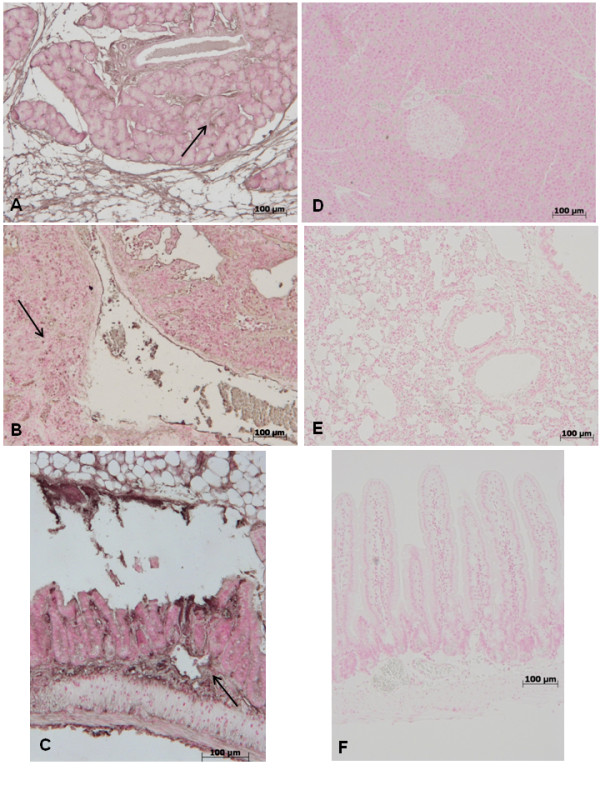
Results: Treatment with GW0742 caused a significant reduction of the peritoneal exudate formation and of the neutrophil infiltration caused by zymosan resulting in a reduction in myeloperoxidase activity. The PPAR-beta/delta agonist, GW0742, at the dose of 0,3 mg/kg in 10% DMSO, also attenuated the multiple organ dysfunction syndrome caused by zymosan. In pancreas, lung and gut, immunohistochemical analysis of some end points of the inflammatory response, such as inducible nitric oxide synthase (iNOS), nitrotyrosine, poly (ADP-ribose) (PAR), TNF- and IL-1as well as FasL, Bax, Bcl-2 and apoptosis, revealed positive staining in sections of tissue obtained from zymosan-injected mice. On the contrary, these parameters were markedly reduced in samples obtained from mice treated with GW0742

Conclusions: In this study, we have shown that GW0742 attenuates the degree of zymosan-induced non-septic shock in mice.


Figures

Figure 1
Figure 1
No histological alteration was observed in the pancreas (a), lung (b) or gut (c) from sham-treated mice Pancreas (d) lung (e) and distal ileum (f) sections from zymosan-administered mice revealed morphological alterations and inflammatory cell infiltration. Pancreas (g) lung (h) and distal ileum (i) from zymosan-administered mice treated with GW0742 demonstrated reduced morphological alterations and inflammatory cell infiltration. Figures are representative of at least 3 experiments performed on different experimental days.
Figure 2
Figure 2
Effect of GW0742-treatment on toxicity score (A), body weight change (B) and mortality (C). Data are means ± SEM of 10 mice for each group. *P < 0.01 vs sham, °P < 0.01 vs zymosan + vehicle.
Figure 3
Figure 3
Effect of GW0742-treatment on inflammatory response in the peritoneal cavity. The increase in volume exudates (A) and peritoneal exudates cell leukocyte counts (B) in peritoneal cavity at 18 h after zymosan was reduced by GW0742 treatment. Data are mean ± standard deviation from n = 10 mice for each group. *P < 0.01 vs sham, °P < 0.01 vs zymosan + vehicle.
Figure 4
Figure 4
Effect of GW0742-treatment on IkB-α degradation and NF-kB p-65 activation. By Western Blot analysis, a basal level of IkB-α was detected in the lung tissue from sham-operated animals, whereas in zymosan-induced mice IkB-α levels were substantially reduced (a, see densitometric analysis a1). Treatment with GW0742 significantly increases the levels of IkB-α, after zymosan injection. Moreover, at 18 h following zymosan-treatment, the levels of NF-kB p-65 subunit protein in the nuclear fractions of the lung tissue were also significant increased compared to the sham-operated mice (b, see densitometric analysis b1). The levels of NF-kB p-65 protein were significantly reduced in the nuclear fractions of the lung tissues from animals that had received GW0742 treatment (b, see densitometric analysis b1). β-actin (a) and Laminin B1 (b) were used as internal control. The result in a1 and b1 are expressed as mean ± S.E. mean from five blots. P < 0.01 vs sham, °P < 0.01 vs zymosan + vehicle.
Figure 5
Figure 5
Effect of GW0742 on plasma tumor necrosis factor alpha (TNF-α) and interleukin-1β (IL-1β) production A substantial increase in TNF-α (A) and IL-1β (B) production was found in tissues collected from zymosan-treated-mice compared to sham mice. Plasma levels of TNF-α and IL-1β were significantly attenuated by the treatment with GW0742, 0.3 mg/Kg 10% DMSO i.p. at 1 and 6 hour after zymosan-injection (A, B, respectively). Data are mean ± standard deviation from n = 10 mice for each group. *P < 0.01 vs sham, °P < 0.01 vs zymosan + vehicle.
Figure 6
Figure 6
and immunohistochemical localization of TNF-α in pancreas, lung and gut. 18 hours following zymosan injection, a positive TNF-α staining was found in pancreas (A), lung (B) and gut (C). There was no detectable immunostaining for TNF-α in pancreas (D), lung (E) and gut (F) of zymosan-treated mice when mice were treated with GW0742. Figures are representative of at least 3 experiments performed on different experimental days.
Figure 7
Figure 7
immunohistochemical localization of IL-1β in pancreas, lung and gut. 18 hours following zymosan injection, a positive IL-1β staining was found in pancreas (A), lung (B) and gut (C). There was no detectable immunostaining for IL-1β in pancreas (D), lung (E) and gut (F) of zymosan-treated mice when mice were treated with GW0742. Figures are representative of at least 3 experiments performed on different experimental days.
Figure 8
Figure 8
Immunohistochemical localization of ICAM-1 in pancreas, lung and gut. 18 hours following zymosan injection, a positive ICAM-1 staining was found in pancreas (A), lung (B) and gut (C). There was no detectable immunostaining for ICAM-1 in pancreas (D), lung (E) and gut (F) of zymosan-treated mice when mice were treated with GW0742. Figures are representative of at least 3 experiments performed on different experimental days.
Figure 9
Figure 9
Immunohistochemical localization of P-Selectin in pancreas, lung and gut. 18 hours following zymosan injection, a positive P-Selectin staining was found in pancreas (A), lung (B) and gut (C). There was no detectable immunostaining for P-Selectin in pancreas (D), lung (E) and gut (F) of zymosan-treated mice when mice were treated with GW0742. Figures are representative of at least 3 experiments performed on different experimental days.
Figure 10
Figure 10
Moreover, MPO activity in lung (A) and gut (B) samples of zymosan-treated-mice was significantly increased in comparision to sham mice (A and B, respectively). Treatment with GW0742 significantly reduced the increase of MPO activity in lung (A) and gut (B). Data are mean ± standard deviation from n = 10 mice for each group. *P < 0.01 vs sham, °P < 0.01 vs zymosan + vehicle.
Figure 11
Figure 11
Effect of GW0742 on peritoneal exudates (A) and plasma nitrate/nitrite levels (B). Nitrate/nitrite levels were significantly increased both in peritoneal exudate and in plasma of zymosan-treated mice in comparison to vehicle group (sham group). GW0742 reduced the zymosan-induced increase of nitrate/nitrite levels in peritoneal exudate (A) and in plasma (B). In addition, by Western Blot analysis the iNOS expression was evaluated in the lung. At 18 h after zymosan administration a significant increase in the iNOS expression was observed in the ileum (Ca, see densitometric analysis a1) compared to the sham-treated mice. GW0742 treatment significantly reduced the iNOS expression (Ca, see densitometric analysis a1). The intensity of bands was measured using a phosphoimager in all the experimental groups. β-actin was used as internal control. A representative blot of lysates obtained from each group is shown, and densitometric analysis of all animals is reported (n = 5 mice from each group). Data are mean ± standard deviation from n = 10 mice for each group. *P < 0.01 vs sham, °P < 0.01 vs zymosan + vehicle.
Figure 12
Figure 12
Immunohistochemical localization of nitrotyrosine in pancreas, lung and gut. 18 h following zymosan injection, a positive nitrotyrosine staining was found in the pancreas (A), lung (B) and gut (C). There was no detectable immunostaining for nitrotyrosine in the pancreas (D), lung (E) and gut (F) of zymosan-administered mice when mice were treated with GW0742. Figure is representative of at least 3 experiments performed on different experimental days.
Figure 13
Figure 13
Immunohistochemical localization of PAR in pancreas, lung and gut. 18 h following zymosan injection, a positive PAR staining was found in the pancreas (A), lung (B) and gut (C). There was no detectable immunostaining for PAR in the pancreas (D), lung (E) and gut (F) of zymosan-administered mice when mice were treated with GW0742. Figure is representative of at least 3 experiments performed on different experimental days.
Figure 14
Figure 14
Effect of GW0742 on Fas-ligand expression. 18 h following zymosan injection, positive FasL staining was found in pancreas (A), lung (B) and gut (C). There was no detectable immunostaining for FasL in pancreas (D), lung (E) and gut (F) of zymosan-administered mice when mice were treated with GW0742. Figures are representative of at least 3 experiments performed on different experimental days.
Figure 15
Figure 15
Effect of GW0742 on TUNEL-like staining. At 18 h after zymosan administration, TUNEL-like staining showed a marked appearance of dark brown apoptotic cells and intercellular apoptotic fragments in pancreatic (A) pulmonary (B) tissue. On the contrary, the number of dark brown cells was significantly reduced in pancreas (C) and lung (D) by the treatment with GW0742. Figures are representative of at least 3 experiments performed on different experimental days.
Figure 16
Figure 16
Western Blot analysis of Bax and Bcl-2 expression. Representative Western Blot of Bax and Bcl-2 levels was realized in pulmonary samples at 18 h after zymosan-injection. At 18 h after zymosan administration, a significant increase in the Bax expression was observed in the lung (A, see densitometric analysis A1) compared to the sham-treated mice, whereas in GW0742-treated mice Bax levels were substantially reduce (A, see densitometric analysis A1). On the contrary, at 18 h after zymosan administration, a decrease in the Bcl-2 expression was observed in the lung (B, see densitometric analysis B1) compared to the sham-treated mice, while Bcl-2 expression was more evident in the pulmonary tissue from zymosan-treated mice that received GW0742 treatment (B, see densitometric analysis B). Data are mean ± standard deviation from n = 10 mice for each group. *P < 0.01 vs sham, °P < 0.01 vs zymosan + vehicle.
Figure 17
Figure 17
Immunohistochemical localization of Bax. Moreover, Bax expression was also evaluated by immunohistochemical analysis in pancreas, lung and gut. 18 hours following zymosan injection, positive Bax staining was found in pancreas (A), lung (B) and gut (C). No positive staining for Bax was detected in pancreas (D), lung (E) and gut (F) of zymosan-treated mice when mice were treated with GW0742. Figures are representative of at least experiments performed on different experimental days.
Figure 18
Figure 18
Immunohistochemical localization of Bcl-2 expression. Moreover, Bcl-2 expression was also evaluated by immunohistochemical analysis in pancreas, lung and gut. At 18 h after zymosan administration, no positive staining for Bcl-2 was observed in pancreas (A), lung (B) and gut (C) from zymosan-treated mice. On the contrary, positive staining for Bcl-2 was observed in pancreas (D), lung (E) and gut (F) from mice treated with GW0742. Figures are representative of at least experiments performed on different experimental days.
Figure 19
Figure 19
Effect of GW0742 on lung injury. Zymosan administration resulted in significant fall in the arterial levels of PaO2 (A), PCO2 (B), pH (C) and HCO3-(D). Administration of GW0742 prevents the lung dysfunctions. Data are means ± SEM of 10 mice for each group. *P < 0.01 vs sham, °P < 0.01 vs zymosan + vehicle.
Figure 20
Figure 20
Effect of GW0742 on liver injury. Administration of zymosan resulted in significantly increased levels of plasma AST (A), ALT (B), bilirubin (C) and alkaline phosphatase (D). GW0742 treatment significantly reduced all these parameters in zymosan treated mice. Data are mean ± standard deviation from n = 10 mice for each group. *P < 0.01 vs sham, °P < 0.01 vs zymosan + vehicle.
Figure 21
Figure 21
Effect of GW0742 on pancreatic and renal injury. Administration of zymosan resulted in significantly increased levels of plasma amylase (A), lipase (B) and creatinine (C). GW0742 treatment significantly decreases all these parameters in zymosan treated mice. Data are mean ± standard deviation from n = 10 mice for each group. *P < 0.01 vs sham, °P < 0.01 vs zymosan + vehicle.

Similar articles

Cited by

References

    1. Volman TJ, Hendriks T, Goris RJ. Zymosan-induced generalized inflammation: experimental studies into mechanisms leading to multiple organ dysfunction syndrome. Shock (Augusta, Ga) 2005;23(4):291–297. doi: 10.1097/01.shk.0000155350.95435.28. - DOI - PubMed
    1. Tenenbaum A, Motro M, Fisman EZ. Dual and pan-peroxisome proliferator-activated receptors (PPAR) co-agonism: the bezafibrate lessons. Cardiovasc Diabetol. 2005;4:14. doi: 10.1186/1475-2840-4-14. - DOI - PMC - PubMed
    1. Sheng L, Ye P, Liu YX, Han CG, Zhang ZY. Peroxisome proliferator-activated receptor beta/delta activation improves angiotensin II-induced cardiac hypertrophy in vitro. Clin Exp Hypertens. 2008;30(2):109–119. doi: 10.1080/10641960801945840. - DOI - PubMed
    1. Braissant O, Foufelle F, Scotto C, Dauca M, Wahli W. Differential expression of peroxisome proliferator-activated receptors (PPARs): tissue distribution of PPAR-alpha, -beta, and -gamma in the adult rat. Endocrinology. 1996;137(1):354–366. doi: 10.1210/en.137.1.354. - DOI - PubMed
    1. Amri EZ, Bonino F, Ailhaud G, Abumrad NA, Grimaldi PA. Cloning of a protein that mediates transcriptional effects of fatty acids in preadipocytes. Homology to peroxisome proliferator-activated receptors. J Biol Chem. 1995;270(5):2367–2371. doi: 10.1074/jbc.270.5.2367. - DOI - PubMed