. 2010 May-Jun;41(3):29.
doi: 10.1051/vetres/2010001. Epub 2010 Jan 13.

Interferon-induced Sus scrofa Mx1 blocks endocytic traffic of incoming influenza A virus particles

Affiliations

Interferon-induced Sus scrofa Mx1 blocks endocytic traffic of incoming influenza A virus particles

Mélanie Palm et al. Vet Res. 2010 May-Jun.

Abstract

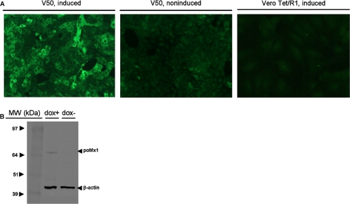
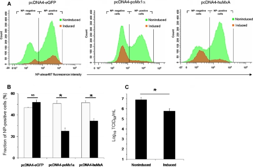
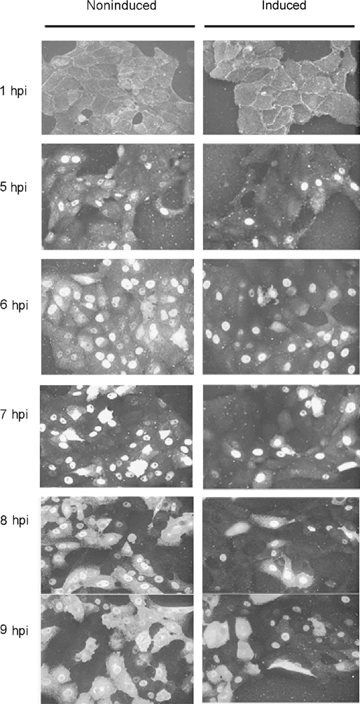
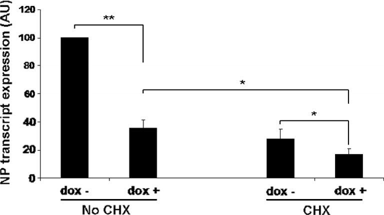
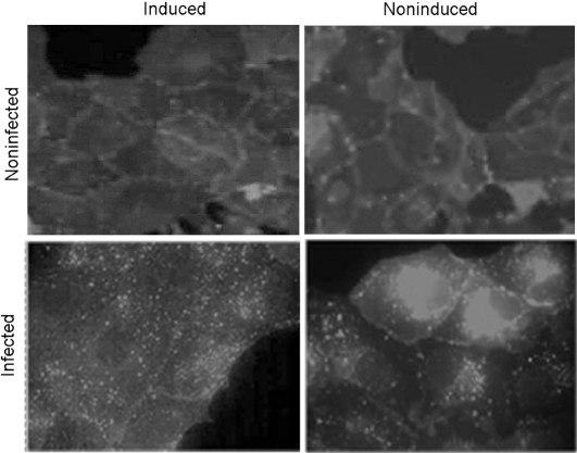
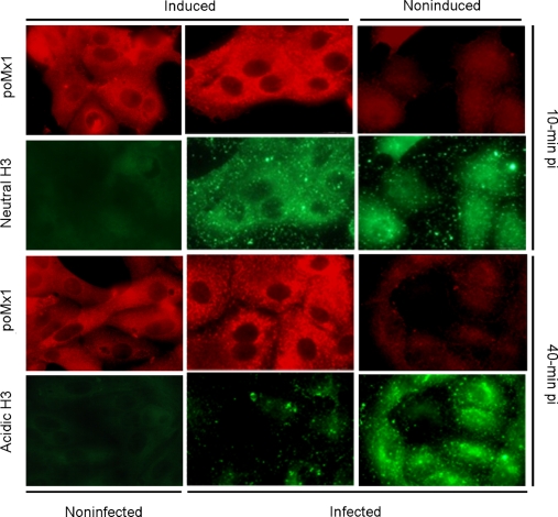
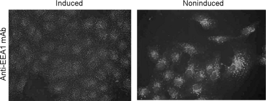
The interferon-induced Mx proteins of vertebrates are dynamin-like GTPases, some isoforms of which can additionally inhibit the life cycle of certain RNA viruses. Here we show that the porcine Mx1 protein (poMx1) inhibits replication of influenza A virus and we attempt to identify the step at which the viral life cycle is blocked. In infected cells expressing poMx1, the level of transcripts encoding the viral nucleoprotein is significantly lower than normal, even when secondary transcription is prevented by exposure to cycloheximide. This reveals that a pretranscriptional block participates to the anti-influenza activity. Binding and internalization of incoming virus particles are normal in the presence of poMx1 but centripetal traffic to the late endosomes is interrupted. Surprisingly but decisively, poMx1 significantly alters binding of early endosome autoantigen 1 to early endosomes and/or early endosome size and spatial distribution. This is compatible with impairment of traffic of the endocytic vesicles to the late endosomes.


Figures

Figure 1.
Figure 1.
Expression of poMx1 in double-transgenic Vero cells examined by fluorescence microscopy and immunoblotting (clone V50). Transgenic cells were induced with 1 μg/mL doxycycline for 24 h. Induced mono-transgenic cells (clone Vero Tet/R1) are presented for control purposes (low background and absence of cross-reactivity with endogenous Cercopithecus aethiops Mx). Clone V50 combined less than 5% spontaneous expression with more than 95% expression and intense cytoplasmic granular staining upon exposure to doxycycline. The Western blot reveals a specific band in the lane for induced cells. The molecular mass corresponding to this band is compatible with that of poMx1 (74 kDa). For indirect immunofluorescence (A), cells were permeabilized and stained by sequential incubation with a rabbit antiserum raised against human MxA (a gift from I. Julkunen, Helsinki, Finland), which had been shown to cross-react with porcine Mx1 [24], and with Alexa 488-conjugated goat anti-rabbit IgG. (B) Immunoblots of 4–12% sodium dodecyl sulfate polyacrylamide gels with total cell proteins extracted from induced (dox +) and non-induced (−) V50 cells (10 μg total protein per lane, as determined by a micro-BCA assay). Immunostaining was done by sequential incubation with a cocktail of the above-mentioned rabbit antiserum and anti-β-actin mAb and a mix of HRP-conjugated anti-rabbit and anti-mouse IgG. Blots were developed by incubation with 3-amino-9-ethylcarbazole. The positions of protein size markers in kilodaltons are indicated (MW). (A color version of this figure is available on line at www.vetres.org.)
Figure 2.
Figure 2.
Porcine Mx1 expression alters influenzavirus NP synthesis (A–B) and production of infectious progeny particles (C). A–B: flow cytometric determination of NP-positive counts in Vero cells transfected with plasmids (pcDNA4) encoding either eGFP (negative control), porcine Mx1 (poMx1α) or human MxA (huMxA, positive control). (A) Example. Cell counts (vertical axis) refer to infected (NP-positive) and noninfected (NP-negative) cells detected in induced (transgene-expressing, dark grey) and non-induced (nonexpressing, light grey) cell populations. (B) Percent NP-positive cells 5 hpi in transgene-expressing (black boxes) and nonexpressing (white) cells. The corresponding plasmids had been transfected 24 h before infection, each population process resulting in a mixed population of expressing and nonexpressing cells. Cells were permeabilized, double immunostained for Mx proteins and virus NP and analysed using the FACS Canto, gating on the forward and side scatter to exclude debris and collecting fluorescences in FL-1 and FL-5. A minimum of 10 000 events were acquired and analyzed with BDFACSDiva software v4.1.1. Each box pair (white/black) represents means ± SD in nonexpressing/expressing cells from 3 independent experiments. Expression of both porcine and human MxA resulted in a significant decrease of NP-positive cell counts at the timepoint studied (5 hpi). (C) Influenza virus yields from stably-transduced porcine Mx1-expressing Vero cell monolayers were significantly decreased after induction of poMx1 expression. Pools of induced (black boxes) and non-induced (white boxes) V50 cells were infected with the H1N1 virus for 48 h (m.o.i. = 0.01). The viral titers in the culture supernatants are plotted, as determined in triplicate by standard median tissue culture infectious dose assays. TCID50: 50% tissue culture infective dose. Plotted values are means ± SD of 3 independent experiments. *Significantly different from the titer retrieved from the corresponding non-induced cells, p < 0.05. (A color version of this figure is available on line at www.vetres.org.)
Figure 3.
Figure 3.
De novo synthesis of H1N1 influenza A virus nucleoprotein (NP) is delayed by porcine Mx1 expression. Production of viral NP by induced and non-induced V50 cells was monitored for 9 h after infection by indirect immunofluorescence, after sequential incubation with the anti-NP mAb from Abcam and Alexa 488-conjugated goat anti-mouse IgG. See text for detailed comparative analysis of NP production scenario in induced and non-induced cells.
Figure 4.
Figure 4.
Porcine Mx1 blocks the influenza A virus life cycle before primary transcription. Cell monolayers were infected with the H1N1 influenza A virus strain for 3 h. Production of transcripts encoding H1N1 influenza A virus NP was quantitated by reverse transcription and real-time PCR in induced and non-induced V50 cells exposed or not, as indicated, to the translation inhibitor cycloheximide (CHX, 100 μg/mL). 18S rRNA was used as an internal control to normalize NP transcript data, and results were expressed percentages of the amount of NP transcripts retrieved from non-induced and nonexposed V50 cells, i.e. the only cells in which the virus life cycle proceeds unhindered. In the absence of CHX, poMx1 expression caused an approximately 65% reduction of NP-encoding mRNA production, and an approximately 40% reduction was still observable after exposure to CHX. Plotted values are means ± SD of triplicate real-time PCR assays, and three independent experiments were performed, with similar results. Fisher’s exact test was used to compare the means and the reported significance levels are p < 0.05 (*) and p < 0.01 (**). AU: arbitrary units. See text for detailed interpretation.
Figure 5.
Figure 5.
Porcine Mx1 expression inhibits the centripetal traffic of endocytic vesicles containing incoming influenza A virus particles. Twenty minutes after addition of the lipophilic dye DiD to the medium, an inoculum drawn from the H1N1 influenza A virus stock and corresponding to a multiplicity of infection of ~ 200 was added to induced and non-induced V50 cells. Infection was stopped 40 min after infection by thorough PBS washings and fixation of cell monolayers with 4% paraformaldehyde. No additional staining was done, so the only visible fluorescence signals are from endocytosed fragments of plasma membrane and viral particles. Forty minutes after infection, non-induced cells displayed endocytosed material concentrated around the cell nuclei. Induced cells, in contrast, reproducibly displayed a diffuse, punctate, “dusty” pattern compatible with a block in the normal centripetal traffic of endocytosed materials.
Figure 6.
Figure 6.
The traffic of incoming viral particles is blocked by poMx1 after internalization but before the acidification-induced conformational change of viral hemagglutinin in the late endosomes. Induced and non-induced cell monolayers were infected with a dilution of the H3N2 influenza A virus stock (m.o.i. ~ 200) and examined 10 min (upper half) and 40 min (lower half) pi after immunostaining of poMx1 (in red) or viral HA (in green). Ten minutes pi, internalized viral particles were detected by labeling the neutral form of the viral HA with N2 mAb. The results are similar for induced and non-induced cells. Forty minutes pi, internalized virus particles having arrived in the perinuclear region were detected by labeling the acidic HA form, this time with the A2 mAb. Non-induced cells show conspicuous staining of acidic HA in the perinuclear region, but induced cells do not. In the latter, traffic to the late endosomes is thus delayed. (A color version of this figure is available on line at www.vetres.org.)
Figure 7.
Figure 7.
Porcine Mx1 expression alters the spatial distribution of structures bearing the early endosome autoantigen 1 (EEA1). Early endosomes were immunostained by sequential incubation with a mAb targeting EEA1 and with an Alexa 488-conjugated goat anti-mouse IgG. Numerous distinct bright granules are visible in non-induced cells, corresponding to early endosomes. Only a diffuse, “dusty” pattern is seen when poMx1 is expressed.

Similar articles

Cited by

References

    1. Apodaca G., Endocytic traffic in polarized epithelial cells: role of actin and microtubule cytoskeleton, Traffic (2001) 2:149–159 - PubMed
    1. Arnheiter H., Haller O., Antiviral state against influenza virus neutralized by microinjection of antibodies to interferon-induced Mx proteins, EMBO J. (1988) 7:1315–1320 - PMC - PubMed
    1. Baise E., Pire G., Leroy M., Gérardin J., Goris N., De Clercq K., et al., Conditional expression of type I interferon-induced bovine Mx1 GTPase in a stable transgenic vero cell line interferes with replication of vesicular stomatitis virus, J. Interferon Cytokine Res. (2004) 24:513–521 - PubMed
    1. Barret T., Wolstenholme A.J., Maby B.W.J., Transcription and replication of influenza virus RNA, Virology (1979) 98:211–225 - PubMed
    1. Bucci C., Parton R.G., Mather I.H., Stunnenberg H., Simons K., Hoflacks B., Zerial M., The small GTPase rab5 functions as a regulatory factor in the early endocytic pathway, Cell (1992) 70:715–728 - PubMed

Publication types

MeSH terms