. 2010 May;92(5):514-29.
doi: 10.1016/j.biochi.2010.02.004. Epub 2010 Feb 16.

Calorimetric and spectroscopic studies of aminoglycoside binding to AT-rich DNA triple helices

Affiliations

Calorimetric and spectroscopic studies of aminoglycoside binding to AT-rich DNA triple helices

Hongjuan Xi et al. Biochimie. 2010 May.

Abstract

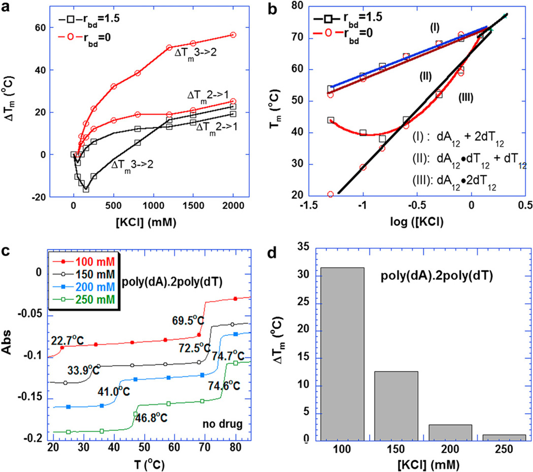
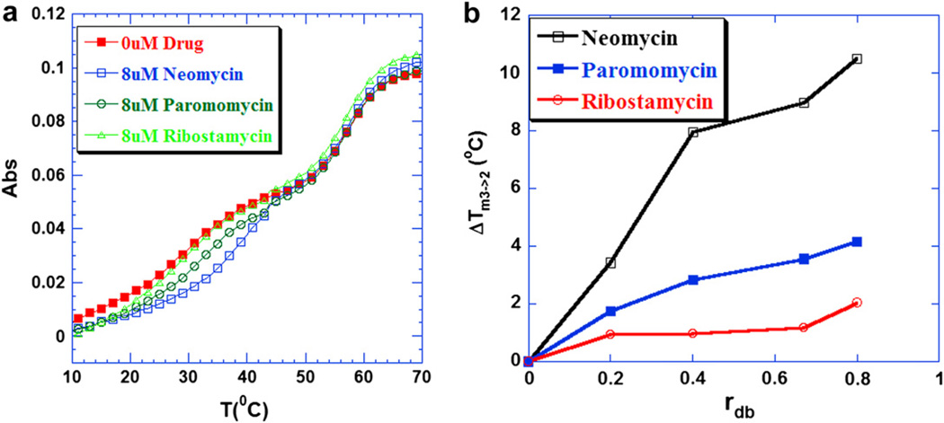
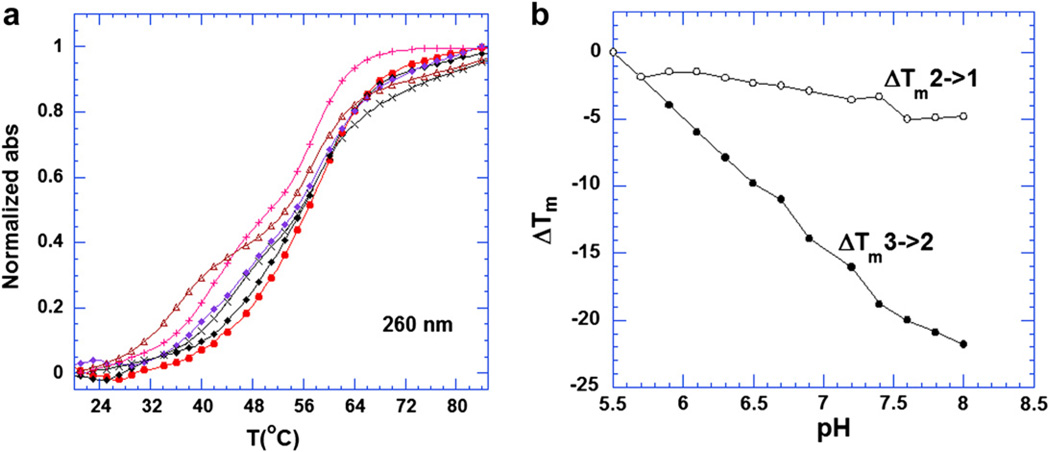
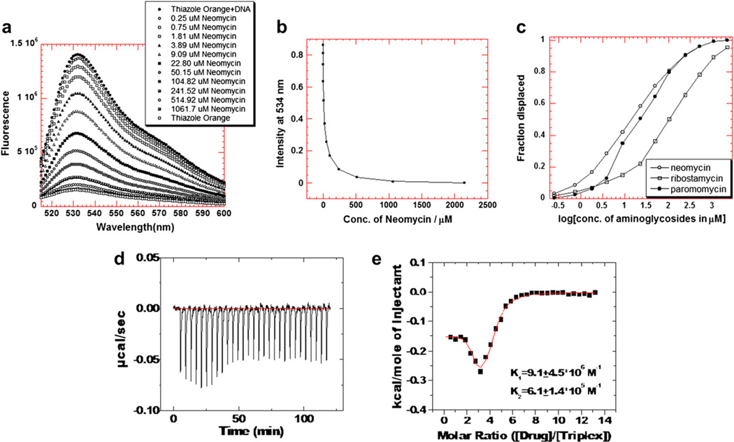
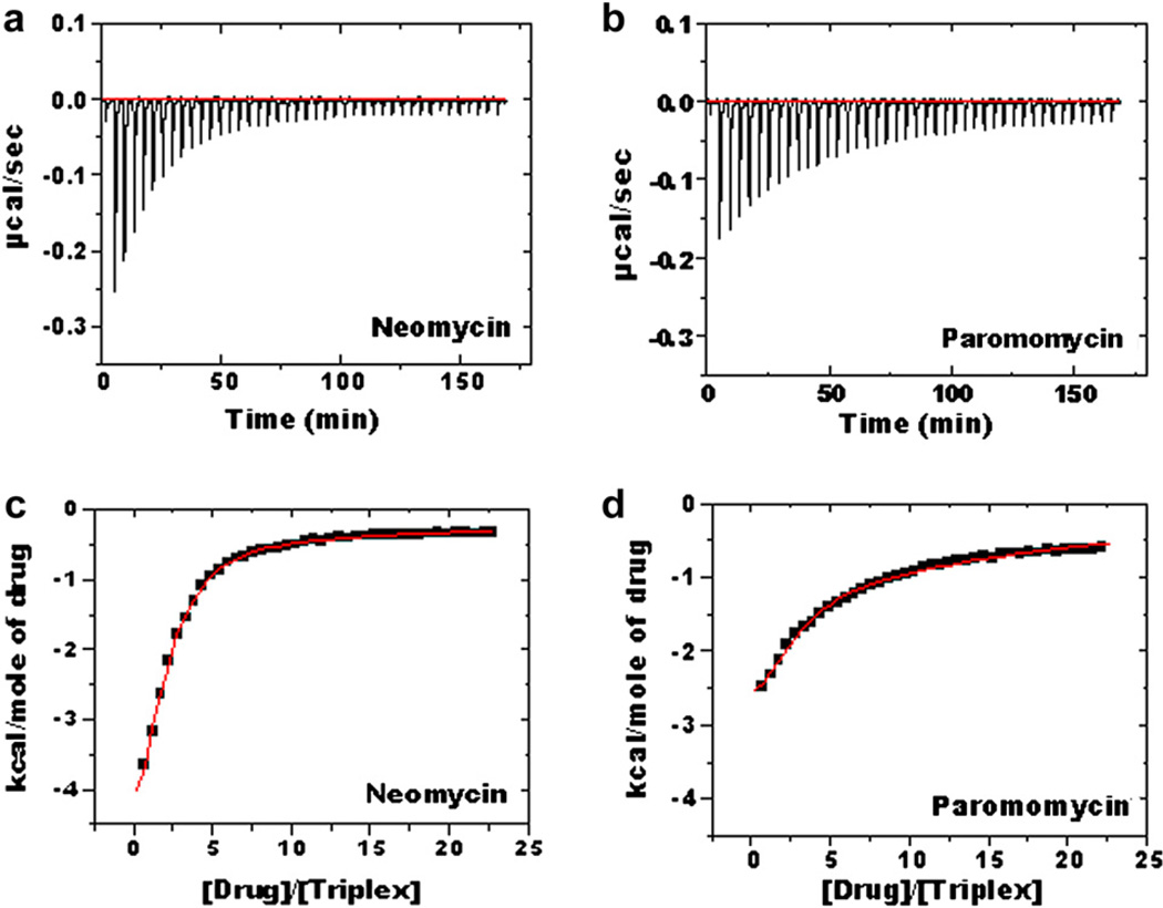
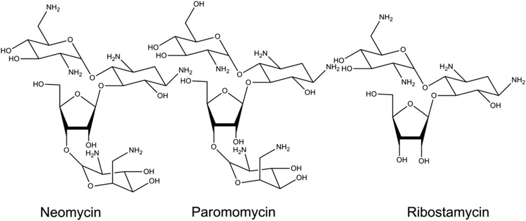
Calorimetric and fluorescence techniques were used to characterize the binding of aminoglycosides-neomycin, paromomycin, and ribostamycin, with 5'-dA(12)-x-dT(12)-x-dT(12)-3' intramolecular DNA triplex (x = hexaethylene glycol) and poly(dA).2poly(dT) triplex. Our results demonstrate the following features: (1) UV thermal analysis reveals that the T(m) for triplex decreases with increasing pH value in the presence of neomycin, while the T(m) for the duplex remains unchanged. (2) The binding affinity of neomycin decreases with increased pH, although there is an increase in observed binding enthalpy. (3) ITC studies conducted in two buffers (sodium cacodylate and MOPS) yield the number of protonated drug amino groups (Deltan) as 0.29 and 0.40 for neomycin and paromomycin interaction with 5'-dA(12)-x-dT(12)-x-dT(12)-3', respectively. (4) The specific heat capacity change (DeltaC(p)) determined by ITC studies is negative, with more negative values at lower salt concentrations. From 100 mM to 250 mM KCl, the DeltaC(p) ranges from -402 to -60 cal/(mol K) for neomycin. At pH 5.5, a more positive DeltaC(p) is observed, with a value of -98 cal/(mol K) at 100 mM KCl. DeltaC(p) is not significantly affected by ionic strength. (5) Salt dependence studies reveal that there are at least three amino groups of neomycin participating in the electrostatic interactions with the triplex. (6) FID studies using thiazole orange were used to derive the AC(50) (aminoglycoside concentration needed to displace 50% of the dye from the triplex) values. Neomycin shows a seven fold higher affinity than paromomycin and eleven fold higher affinity than ribostamycin at pH 6.8. (7) Modeling studies, consistent with UV and ITC results, show the importance of an additional positive charge in triplex recognition by neomycin. The modeling and thermodynamic studies indicate that neomycin binding to the DNA triplex depends upon significant contributions from charge as well as shape complementarity of the drug to the DNA triplex Watson-Hoogsteen groove.
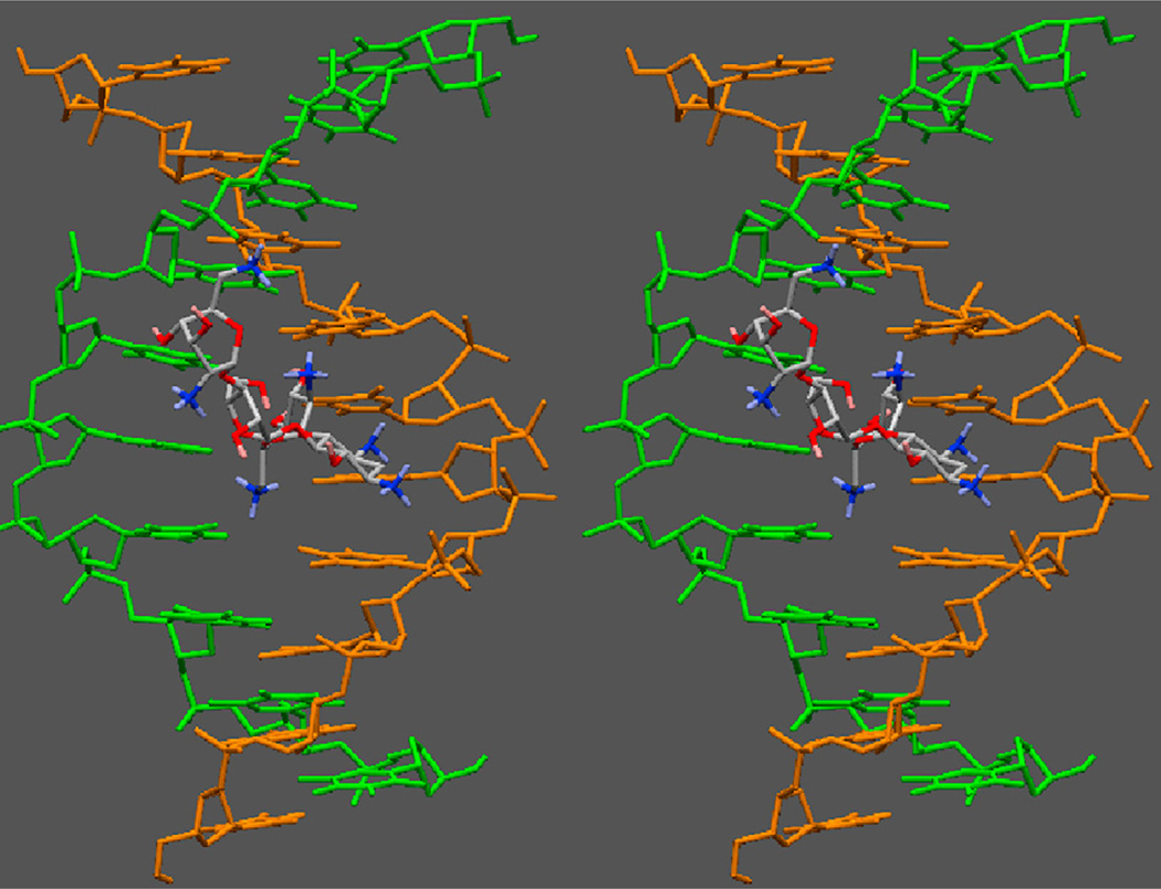
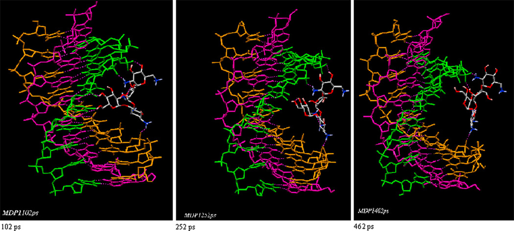

Figures

Fig. 1
Fig. 1
(a) Plots of ΔTm3→2 and ΔTm2→1 of the 5′-dA12-x-dT12-x-dT12-3′ triplex as a function of increasing KCl with or without 8 mM neomycin. (b) Phase diagram of Tm as a function of log [K+] for 5′-dA12-x-dT12-x-dT12-3′ triplex with or without 8 µM neomycin. The data points represent the melting temperature. Each phase labeled as I, II, and III refers to the single strand, duplex and triplex structural states, respectively, that DNA adopts. (c) UV melting profile of poly(dA).2poly(dT) at various KCl. (d) A bar graph of triplex melting temperature increase (ΔTm) of poly(dA).2poly(dT) at saturated amount of neomycin. Buffer conditions: 10 mM sodium cacodylate, 0.5 mM EDTA, and pH 6.8. DNA triplex concentration was 1 µM/strand for oligomer and 15 µM/base triplet for polymer.
Fig. 2
Fig. 2
(a) UV melting profiles of 5′-dA12-x-dT12-x-dT12-3′ triplex at 260 nm in the presence of aminoglycosides. From left to right, the UV melting profiles correspond to 0 µM drug, 8 µM ribostamycin, paromomycin and neomycin respectively. (b) A plot of ΔTm3→2 of 5′-dA12-x-dT12-x-dT12-3′ triplex as a function of increasing rdb values [rdb = drug/base triplet ratio]. Buffer conditions: 10 mM sodium cacodylate, 0.5 mM EDTA, 100 mM KCl, and pH 7.2. DNA triplex concentration was 1 µM/strand.
Fig. 3
Fig. 3
(a) UV melting profiles at 260 nm of the 5′-dA12-x-dT12-x-dT12-3′ triplex in the presence of 8 µM neomycin (rdb = 0.75). From left to right, the pH values of solutions were 7.4, 6.9, 6.7, 6.3, 5.9 and 5.5, respectively. (b) Plots of the ΔTm3→2 and ΔTm2→1 of the 5′-dA12-x-dT12-x-dT12-3′ triplex as a function of increasing pH value in the presence of 8 µM neomycin (rdb = 0.75). The Tm for the triplex was determined from the profiles at 280 nm ΔTm = Tm(any pH)-Tm(5.5). All the buffer solutions contained 10 mM sodium cacodylate, 0.5 mM EDTA and 100 mM KCl. DNA triplex concentration was 1 µM in strand.
Fig. 4
Fig. 4
(a–c). A graphical representation of thiazole orange displacement assay (a) A raw emission data of 1.25 µM thiazole orange upon excitation at 534 nm with buffer only (open circles) and after addition of 100 nM/strand triplex deoxynucleotide hairpin 5′-dA12-x-dT12-x-dT12-3′ (black circles). Neomycin was then titrated from 0.25 µM to 1.06 mM. (b) The decrease in the fluorescence intensity of the complex (DNA-thiazole orange) upon addition of neomycin aliquots. (c) Assuming a linear relationship between the changes in fluorescence intensity with the fraction of thiazole orange displaced results in S-shaped binding isotherm. This graph allows the determination of concentration of ligands needed to displace half of the thiazole orange from triplex deoxynucleotide hairpin 5′-dA12-x-dT12-x-dT12-3′. (d–e) ITC titration of neomycin with intrramolecular triplex at pH 5.5. (d) ITC profiles of neomycin binding with 5′-dA12-x-dT12-x-dT12-3′ triplex. (e) Corrected injection heat as a function of [drug]/[triplex] ratio. The data points represent the experimental injection heat and the solid lines were corresponding to the calculated fits of the data using a model with two sets of binding sites (Origin 5.0). Buffer condition: 10 mM sodium cacodylate, 0.5 mM EDTA, 150 mM KCl, pH 5.5. T= 10 °C.
Fig. 5
Fig. 5
ITC studies of intramolecular triplex 5′-dA12-x-dT12-x-dT12-3′ with neomycin (a) and paromomycin (b) in 10 mM cacodylate, 0.5 mM EDTA, 150 mM KCl, pH 6.8; T= 10 °C [DNA] = 4 µM/strand, [drug] = 600 µM (c–d) Corrected injection heat as a function of [drug]/[triplex] ratio. The data points represent the experimental injection heat and the solid lines correspond to the calculated fits of the data by using a model with two binding sites (Origin 5.0).
Fig. 6
Fig. 6
UV melting profiles of poly(dA).2poly(dT) triplex in the absence (1) and presence of neomycin (2) with rbd 8.8. (b) A plot of ITC derived binding enthalpies vs. corresponding temperatures. The slope reflects the heat capacity change ΔCp. (c) DSC melting profile of poly(dA).2poly(dT). Buffer condition: 10 mM sodium cacodylate, 0.5 mM EDTA, 100 mM KCl, and pH 5.5.
Fig. 7
Fig. 7
Salt dependence of the neomycin binding with poly(dA)-2poly(dT) triplex in 10 mM sodium cacodylate, 0.5 mM EDTA and pH 5.5. T= 10 °C. The experimental data were fit with linear regression and the solid line reflects the resulting curve fit.
Fig. 8
Fig. 8
Stereo view of neomycin bound to the W–H groove of the DNA triplex. Only the two pyrimidine strands are shown for clarity. Neomycin occupies 6–7 base-pairs per triplex.
Fig. 9
Fig. 9
A 500 ps MD simulation of paromomycin bound to the W–H groove of the DNA triplex, with ring I inside the groove. Three snapshots show the dissociation of paromomycin with time. Dissociation of ring I drives the molecule outside the groove.
Scheme 1
Scheme 1
Hydrogen bonds formed in pyrimidine.purine-pyrimidine triplets (pyrimidine motif) and purine.purine-pyrimidine triplets (purine motif).
Scheme 2
Scheme 2
Structures of the aminoglycosides used in the study.

Similar articles

Cited by

References

    1. Maher LJ. Prospects for the therapeutic use of antigene oligonucleotides. Cancer Invest. 1996;14:66–82. - PubMed
    1. Giovannangeli C, Helene C. Progress in developments of triplex-based strategies antisense. Nucleic Acid Drug Dev. 1997;7:413–421. - PubMed
    1. Praseuth D, Guieysse-Peugeot A, Helene C. Triple helix formation and the antigene strategy for sequence-specific control of gene expression. Biochem. Biophys. Res. Commun. 1999;1489:181–206. - PubMed
    1. Mirkin SM, Lyamichev VI, Drushlyak KN, Dobrynin VN, Filippov SA, Frank-Kamenetskii M. DNA H form reuires a homopurine-homopyrimidine mirror repeat. Nature. 1987;330:495–497. - PubMed
    1. Maher LJI, Wold B, Dervan PB. Inhibition of DNA binding proteins by oligonucleotide-directed triple helix formation. Science. 1989;245:725–730. - PubMed

Publication types

LinkOut - more resources