. 2010 Apr 16;285(16):11913-21.
doi: 10.1074/jbc.M109.083238. Epub 2010 Feb 18.

Nucleosome formation activity of human somatic nuclear autoantigenic sperm protein (sNASP)

Affiliations

Nucleosome formation activity of human somatic nuclear autoantigenic sperm protein (sNASP)

Akihisa Osakabe et al. J Biol Chem. .

Abstract

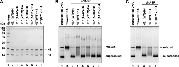
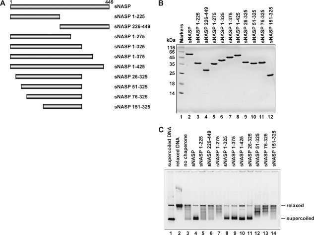
NASP (nuclear autoantigenic sperm protein) is a member of the N1/N2 family, which is widely conserved among eukaryotes. Human NASP reportedly prefers to bind to histones H3.H4 and the linker histone H1, as compared with H2A.H2B, and is anticipated to function as an H3.H4 chaperone for nucleosome assembly. However, the direct nucleosome assembly activity of human NASP has not been reported so far. In humans, two spliced isoforms, somatic and testicular NASPs (sNASP and tNASP, respectively) were identified. In the present study we purified human sNASP and found that sNASP efficiently promoted the assembly of nucleosomes containing the conventional H3.1, H3.2, H3.3, or centromere-specific CENP-A. On the other hand, sNASP inefficiently promoted nucleosome assembly with H3T, a testis-specific H3 variant. Mutational analyses revealed that the Met-71 residue of H3T is responsible for this inefficient nucleosome formation by sNASP. Tetrasomes, composed of the H3.H4 tetramer and DNA without H2A.H2B, were efficiently formed by the sNASP-mediated nucleosome-assembly reaction. A deletion analysis of sNASP revealed that the central region, amino acid residues 26-325, of sNASP is responsible for nucleosome assembly in vitro. These experiments are the first demonstration that human NASP directly promotes nucleosome assembly and provide compelling evidence that sNASP is a bona fide histone chaperone for H3.H4.


Figures

FIGURE 1.
FIGURE 1.
Purification of human sNASP. A, proteins from each purification step were analyzed by 15% SDS-PAGE with Coomassie Brilliant Blue staining. Lane 1 indicates the molecular mass markers. Lanes 2 and 3 are the whole cell lysates before and after induction with isopropyl-β-d-thiogalactopyranoside (IPTG), respectively. Lane 4 indicates the sample from the peak Ni-NTA-agarose fraction. B, an 8% SDS-PAGE image of the Ni-NTA-agarose fraction before (lane 2) and after (lane 3) the removal of the His6 tag by thrombin treatment is shown. C, SDS-PAGE analysis of purified sNASP lacking the His6 tag.
FIGURE 2.
FIGURE 2.
Nucleosome assembly activity of sNASP. A, electrophoretic mobility shift assay for histone H3·H4 binding by sNASP is shown. hNap1 (2.3 μg) or sNASP (2.4 μg) was incubated with different amounts of H3.1·H4, and the hNap1·H3.1·H4 or sNASP·H3.1·H4 complex was detected by non-denaturing 5% PAGE with Coomassie Brilliant Blue staining. The amounts of H3.1·H4 were 0 μg (lanes 1 and 7), 0.375 μg (lanes 2 and 8), 0.75 μg (lanes 3 and 9), 1.5 μg (lanes 4 and 10), 2.25 μg (lanes 5 and 11), and 3 μg (lanes 6 and 12). B, nucleosome assembly assay is shown. A 195-bp 5 S DNA fragment was incubated with histones, and the nucleosome assembly reactions were performed with hNap1 (lane 2) or sNASP (lane 3). Lane 1 indicates a negative control experiment without histone chaperone. The samples were analyzed by non-denaturing 6% PAGE. C, shown is an MNase assay. The nucleosome assembly reactions were performed with hNap1 (lanes 7–11) or sNASP (lanes 12–16). For negative control experiments, the reactions were also performed without histone chaperone (lanes 2–6). The samples were then treated with MNase, and the resulting DNA fragments were analyzed by non-denaturing 10% PAGE. The amounts of MNase were 0.8 unit (lanes 2, 7, and 12), 0.4 unit (lanes 3, 8, and 13), 0.2 unit (lanes 4, 9, and 14), 0.1 unit (lanes 5, 10, and 15), and 0 unit (lanes 6, 11, and 16). NCP indicates DNA fragments tightly wrapped around histone octamers.
FIGURE 3.
FIGURE 3.
Topological assay for nucleosome assembly by sNASP. A, relaxed ϕX174 DNA (10 ng/μl), which was previously treated with wheat germ topoisomerase I (lane 2), was incubated with hNap1 (lanes 4–6) or sNASP (lanes 9–11) in the presence of core histones. The reaction products were then analyzed by 1% agarose gel electrophoresis in 1 × TAE buffer. Lanes 3 and 8 indicate negative control experiments without histone chaperone in the presence of core histones. Lanes 7 and 12 indicate the other negative control experiments without core histones in the presence of hNap1 (1 μm) or sNASP (1 μm). The concentrations of hNap1 and sNASP were 0.25 μm (lanes 4 and 9), 0.5 μm (lanes 5 and 10), and 1 μm (lanes 6 and 11). B, tetrasome assembly is shown. Reactions were conducted as described for the experiments shown in panel A, except H2A·H2B or H3.1·H4 were used instead of the four core histones, H2A·H2B/H3.1·H4. Lanes 3 and 8 indicate negative control experiments without histone chaperone in the presence of H2A·H2B or H3.1·H4, respectively. Lanes 5, 7, 10, and 12 indicate the other negative control experiments without core histones in the presence of either hNap1 (1 μm) or sNASP (1 μm). Lanes 3, 4, and 6 represent experiments with H2A·H2B, and lanes 8, 9, and 11 represent experiments with H3.1·H4.
FIGURE 4.
FIGURE 4.
Interactions between sNASP and human H3 variants. A, shown is a 16% SDS-PAGE analysis of purified human H3 variants complexed with H4. B, shown is an electrophoretic mobility shift assay. sNASP (2.4 μg) was incubated with different amounts of H3.1·H4 (lanes 2–5), H3.2·H4 (lanes 7–10), H3.3·H4 (lanes 12–15), H3T·H4 (lanes 17–20), CENP-A·H4 (lanes 22–25), and H2A·H2B (lanes 27–30), and the complexes were detected by non-denaturing 5% PAGE with Coomassie Brilliant Blue staining. The amounts of histones were 0 μg (lanes 1, 6, 11, 16, 21, and 26), 0.75 μg (lanes 2, 7, 12, 17, 22, and 27), 1.5 μg (lanes 3, 8, 13, 18, 23, and 28), 2.25 μg (lanes 4, 9, 14, 19, 24, and 29), and 3 μg (lanes 5, 10, 15, 20, 25, and 30). C, shown is a graphic representation of the sNASP binding to H3.1·H4, H3.2·H4, H3.3·H4, H3T·H4, and CENP-A·H4. The relative amounts of sNASP in the complex with H3.1·H4 (closed circles), H3.2·H4 (closed triangles), H3.3·H4 (closed squares), H3T·H4 (open circles), and CENP-A·H4 (open triangles) are plotted as the averages of three independent experiments with the S.D. values.
FIGURE 5.
FIGURE 5.
Nucleosome assembly with human H3 variants by sNASP. A, nucleosome assembly with H3 variants by sNASP is shown. The topological assay was employed. Relaxed ϕX174 DNA (10 ng/μl), which was previously treated with wheat germ topoisomerase I (lane 2), was incubated with hNap1 (lanes 4, 7, 10, 13, and 16) or sNASP (lanes 5, 8, 11, 14, and 17) in the presence of core histones. Lanes 3–5, 6–8, 9–11, 12–14, and 15–17 indicate experiments with H3.1·H4, H3.2·H4, H3.3·H4, H3T·H4, and CENP-A·H4, respectively. The reaction products were then analyzed by 1% agarose gel electrophoresis in 1 × TAE buffer. Lanes 3, 6, 9, 12, and 15 indicate negative control experiments without histone chaperone in the presence of core histones. The concentration of hNap1 or sNASP was 1 μm. B, the H3.1 and H3T nucleosomes reconstituted by salt dialysis were fractionated using a Prepcell apparatus and were analyzed by non-denaturing 6% PAGE. C, histone compositions of the purified H3.1 and H3T nucleosomes were analyzed by 18% SDS-PAGE. D, shown are protein titration experiments. The reactions were conducted as described in panel A. Lanes 3–6, 7–10, 11–14, 15–18, and 19–22 indicate experiments with H3.1·H4, H3.2·H4, H3.3·H4, H3T·H4, and CENP-A·H4, respectively. The concentrations of sNASP were 0 μm (lanes 3, 7, 11, 15, and 19), 0.1 μm (lanes 4, 8, 12, 16, and 20), 0.2 μm (lanes 5, 9, 13, 17, and 21), and 0.4 μm (lanes 6, 10, 14, 18, and 22). Lane 23 represents a negative control experiment without core histones. E, shown is deposition of H3.1·H4 or H3T·H4 from the sNASP·H3.1·H4 or sNASP·H3T·H4 complex onto DNA, analyzed by non-denaturing 5% PAGE with Coomassie Brilliant Blue staining. sNASP (2.4 μg) was incubated without or with H3.1·H4 (1 μg) or H3T·H4 (1.5 μg) to form the complex; about half of the sNASP remained free under these conditions. After the incubation with supercoiled DNA, the samples were analyzed. The amounts of competitor DNA were 0, 10, 20, 30, 40, 50, 60, 70, 80, 90, and 100 ng. The amounts of sNASP released from sNASP·H3.1·H4 (closed circles) or sNASP·H3T·H4 complex (open circles) are plotted as the averages of three independent experiments with the S.D. values.
FIGURE 6.
FIGURE 6.
Mutational analyses of the amino acid residue(s) required for H3T incorporation into nucleosomes. A, shown is SDS-PAGE analysis of the H3.1 and H3T mutants complexed with H4. Lane 1, molecular mass markers. Lanes 2–6, H3.1 mutants complexed with H4; lane 2, H3.1(WT); lane 3, H3.1(A24V); lane 4; H3.1(V71M); lane 5; H3.1(A98S); lane 6; H3.1(A111V). Lanes 7 and 8, H3T mutants complexed with H4; lane 7, H3T(WT), lane 8, H3T(M71V). B, nucleosome reconstitution with the H3.1 mutants by sNASP is shown. The topological assay was employed. Relaxed ϕX174 DNA (10 ng/μl), which was previously treated with wheat germ topoisomerase I (lane 2), was incubated with sNASP in the presence of core histones. Lanes 3–8 indicate experiments with H3.1(WT)·H4, H3T(WT)·H4, H3.1(A24V)·H4, H3.1(V71M)·H4, H3.1(A98S)·H4, and H3.1(A111V)·H4, respectively. The reaction products were then analyzed by 1% agarose gel electrophoresis in 1 × TAE buffer. The sNASP concentration was 1 μm. C, nucleosome reconstitution with the H3T(M71V) mutant by sNASP is shown. The topological assay was employed. Relaxed ϕX174 DNA (10 ng/μl), which was previously treated with wheat germ topoisomerase I (lane 2), was incubated with sNASP in the presence of core histones. Lanes 3–5 indicate experiments with H3.1(WT)·H4, H3T(WT)·H4, and H3T(M71V)·H4, respectively.
FIGURE 7.
FIGURE 7.
Deletion analysis of sNASP. A, shown is a schematic representation of the sNASP fragments. B, shown is a 15% SDS-PAGE analysis of the purified sNASP fragments (lanes 3–12). These sNASP fragments lack the His6 tag. Lane 1 indicates the molecular mass markers. Lane 2 represents full-length sNASP. C, nucleosome assembly by the sNASP fragments is shown. Relaxed ϕX174 DNA (10 ng/μl), which was previously treated with wheat germ topoisomerase I (lane 2), was incubated with sNASP or the sNASP fragments in the presence of core histones. Lane 3 indicates a negative control experiment without sNASP or the sNASP fragments in the presence of core histones. The reaction products were then analyzed by 1% agarose gel electrophoresis in 1 × TAE buffer. The concentrations of sNASP and the sNASP fragments were 1 μm.

Similar articles

Cited by

References

    1. Luger K., Mäder A. W., Richmond R. K., Sargent D. F., Richmond T. J. (1997) Nature 389, 251–260 - PubMed
    1. Arents G., Burlingame R. W., Wang B. C., Love W. E., Moudrianakis E. N. (1991) Proc. Natl. Acad. Sci. U.S.A. 88, 10148–10152 - PMC - PubMed
    1. Akey C. W., Luger K. (2003) Curr. Opin. Struct. Biol. 13, 6–14 - PubMed
    1. Loyola A., Almouzni G. (2004) Biochim. Biophys. Acta 1677, 3–11 - PubMed
    1. Henikoff S., Ahmad K. (2005) Annu. Rev. Cell Dev. Biol. 21, 133–153 - PubMed

Publication types

MeSH terms

LinkOut - more resources