. 2010 Apr 23;285(17):13201-10.
doi: 10.1074/jbc.M109.058669. Epub 2010 Feb 18.

Activating transcription factor 3 activates p53 by preventing E6-associated protein from binding to E6

Affiliations

Activating transcription factor 3 activates p53 by preventing E6-associated protein from binding to E6

Hongbo Wang et al. J Biol Chem. .

Abstract

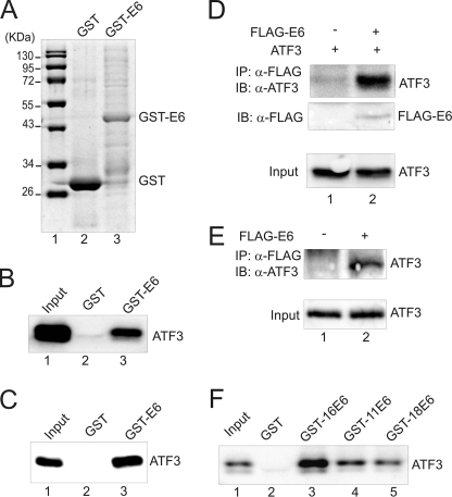
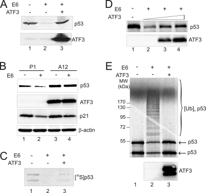
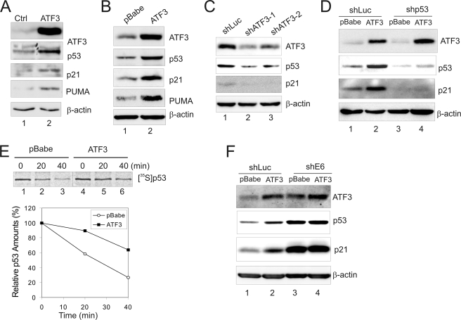
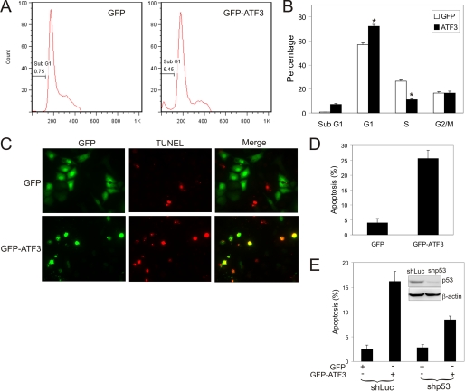
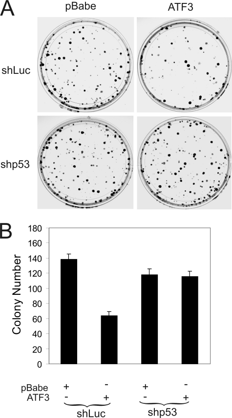
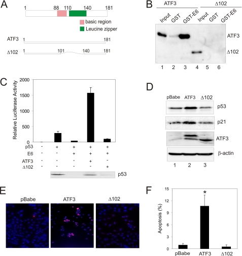
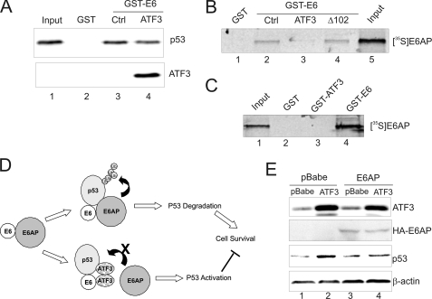
Genomic integration of human papillomavirus (HPV) DNA accounts for more than 90% of cervical cancers. High-risk genital HPVs encode E6 proteins that can interact with a cellular ubiquitin ligase E6-associated protein (E6AP) and target the tumor suppressor p53 for ubiquitin-mediated proteolysis. Currently, how this critical event is regulated is largely unknown. Here we report that activating transcription factor 3 (ATF3), a broad DNA damage sensor whose expression is frequently downregulated in cervical cancer, interacted with E6 and prevented p53 from ubiquitination and degradation mediated by the viral protein. Consistent with its role as a potent E6 antagonist, ATF3 expressed enforcedly in HPV-positive SiHa cells activated p53, leading to expression of p53-target genes (e.g. p21 and PUMA), cell cycle arrest and apoptotic cell death. The leucine zipper domain of ATF3 appears indispensable for these effects as an ATF3 mutant lacking this domain failed to interact with E6 and activate p53 in the cervical cancer cells. The prevention of p53 degradation was unlikely caused by binding of ATF3 to the tumor suppressor, but rather was a consequence of disruption of the E6-E6AP interaction by ATF3. These results indicate that ATF3 plays a key role in a mechanism defending against HPV-induced carcinogenesis, and could serve as a novel therapeutic target for HPV-positive cancers.


Figures

FIGURE 1.
FIGURE 1.
ATF3 directly binds to E6. A, GST-E6 and GST proteins eluted from glutathione-agarose were subjected to SDS-PAGE and stained with Coomassie Blue. B, immobilized GST-E6 or GST protein was incubated with in vitro-translated ATF3 protein at 4 °C overnight. After extensive washes, bound proteins were eluted and subjected to immunoblotting with an ATF3 antibody. Lane 1 represents 10% of total input protein. C, immobilized GST-E6 or GST was incubated with 200 ng of purified ATF3 protein followed by immunoblotting to visualize bound proteins. The input lane represents 10% of total input protein. D, ATF3 was expressed in H1299 cells with or without FLAG-E6 by transfections. Cell lysates were immunoprecipitated by agarose conjugated with an anti-FLAG antibody, and bound proteins were eluted for immunoblotting as indicated. E, HCT116 p53−/− cells were transfected with the plasmid encoding FLAG-E6. After treated with 25 μm MG132, cells were subjected to immunoprecipitations as in D to examine the interaction of FLAG-E6 with the endogenous ATF3 protein. F, GST-E6 proteins derived from HPV11 (GST-11E6), HPV16 (GST-16E6), and HPV18 (GST-18E6) were immobilized on glutathione-agarose, and incubated with in vitro-translated ATF3 proteins for GST-pulldown assays as in B.
FIGURE 2.
FIGURE 2.
ATF3 prevents p53 from E6-mediated degradation and ubiquitination. A, H1299 cells were co-transfected with p53, E6, RSV-Luc, and ATF3 as indicated. 48 h after transfections, cell lysates were normalized against luciferase activity (as transfection efficiency control) and subjected to immunoblotting for p53 and ATF3 expression. B, HT1080 cells stably expressing ATF3 (A12) or its empty vector (P1) (24) were transfected with E6 for 2 days, and lysed for immunoblotting analysis. C, E6 protein was preincubated with (lane 3) or without ATF3 (lane 2) at 4 °C for 30 min, and then added into a reaction mixture containing 35S-labeled p53. The reactions were terminated after 60 min, and p53 was visualized by autography. D, E6 was preincubated with increasing amounts of ATF3, and mixed with in vitro-translated p53 for 60 min. The p53 levels were measured by immunoblotting using the DO-1 antibody. E, E6 protein preincubated with (lane 3) or without ATF3 (lane 2) was incubated with 35S-labeled p53 in the presence of 20 μg of methylated ubiquitin for 60 min followed by SDS-PAGE and autography.
FIGURE 3.
FIGURE 3.
ATF3 activates p53 in HPV-positive cancer cells. A, SiHa cells were transfected with GFP (lane 1) or GFP-IRES-ATF3 (lane 2) for 2 days. The sorted GFP-positive cells were lysed for immunoblotting. B, SiHa cells were infected with retroviruses expressing ATF3 (lane 2) or its vector pBabe (lane 1) for 2 days followed by immunoblotting. C, SiHa cells were infected with lentiviruses expressing two independent shRNA targeting ATF3 (lanes 2 and 3) or luciferase (lane 1) for 3 days followed by immunoblotting. D, SiHa cells were sequentially infected with lentiviruses expressing shRNA specific to p53 (shp53) or luciferase (shLuc), and retroviruses expressing ATF3 or its vector as indicated. Infected cells were lysed and subjected to immunoblotting. E, SiHa cells infected with retroviruses expressing ATF3 or pBabe were labeled with Tran[35S] for 1 h and lysed at indicated time. p53 was immunoprecipitated with the DO-1 antibody and visualized by autography. The graph shows results of densitometric analysis. F, SiHa cells were infected sequentially with retroviruses expressing ATF3 and E6-targeted shRNA, and then lysed for immunoblotting as indicated.
FIGURE 4.
FIGURE 4.
ATF3 induces SiHa cells to cell cycle arrest and apoptosis as a consequence of p53 activation. A and B, SiHa cells were transfected with GFP or GFP-ATF3 for 2 days, and stained with PI. GFP-positive cells were subjected to cell cycle analysis using the FlowJo software. *, p < 0.001. C and D, SiHa cells were transfected with GFP or GFP-ATF3 for 3 days, and subjected to TUNEL staining. Quantitation was performed by counting at least 300 GFP-positive cells. E, SiHa cells were infected with Lentiviruses expressing shp53 or shLuc for 3 days, followed by transfections with GFP or GFP-ATF3 for 3 days. The cells were stained with TUNEL and counted as C. The inset shows an immunoblot for p53 expression.
FIGURE 5.
FIGURE 5.
ATF3 decreases the growth potential of HPV-positive cells. SiHa cells infected with shp53- or shLuc-expressing Lentiviruses were further infected with retroviruses expressing ATF3 or its vector pBabe for 2 days. The infected cells (200/well) were plated into 6-well plates, and cultured for 14 days. The colonies were stained with crystal violet (A) and counted (B).
FIGURE 6.
FIGURE 6.
The ATF3 Zip domain is required for E6 binding and p53 activation in HPV-positive cells. A, schematic representation of the full-length ATF3 protein and the mutant protein lacking the Zip domain (Δ102) purified from bacterial culture using Ni2+-nitrilotriacetic acid-agarose. The recombinant proteins contain a c-Myc tag that can be used for detection of the mutant protein by immunoblotting. B, purified ATF3 and Δ102 proteins were incubated with immobilized GST-E6 or E6, and subjected to GST-pulldown assays as in Fig. 1C. The bound proteins were detected with an anti-c-Myc antibody. The input lane represents 10% of total input protein. C, H1299 cells were co-transfected with a p53 luciferase reporter, pRL-TK, p53, E6, ATF3, or Δ102 as indicated. Dual luciferase assays were performed to measure the p53 transcription activity. The immunoblot shows p53 expression in the transfected cells. D, SiHa cells were infected with retroviruses expressing ATF3, Δ102, or pBabe, and subjected to immunoblotting for detection of p53 and p21 expression. E and F, SiHa cells were infected with pBabe, ATF3, or Δ102, and then subjected to TUNEL staining followed by counterstaining with DAPI. Representative, merge images were shown in E. At least 300 cells were counted to calculate percentages of apoptotic cells (F). *, p < 0.001 compared with the pBabe group.
FIGURE 7.
FIGURE 7.
ATF3 prevents E6AP from binding to E6. A, in vitro-translated p53 protein was preincubated with BSA (lane 3) or ATF3 (lane 4) before incubated with immobilized GST-E6 or GST. Bound proteins were visualized by immunoblotting. The input lane represents 10% of total input protein. B, immobilized E6 protein was preincubated with BSA (lane 2), ATF3 (lane 3), or Δ102 (lane 4), and then incubated with 3 μl of 35S-labeled E6AP for 4 h. Bound proteins were visualized by autography. C, 3 μl of 35S-labeled E6AP was incubated with immobilized GST, GST-ATF3, or GST-E6, followed by autography to visualize bound proteins. D, schematic representation of a model in which ATF3 competes with E6AP for binding to E6 thereby blocking p53 ubiquitination catalyzed by the E3 ubiquitin ligase. E, SiHa cells were sequentially infected with retroviruses expressing HA-E6AP and ATF3 for 2 days and lysed for immunoblotting to determine the p53 level.

Similar articles

Cited by

References

    1. zur Hausen H. (2000) J. Natl. Cancer Inst. 92, 690–698 - PubMed
    1. Schiffman M., Castle P. E., Jeronimo J., Rodriguez A. C., Wacholder S. (2007) Lancet 370, 890–907 - PubMed
    1. Scheffner M., Werness B., Huibregtse J. M., Levine A. J., Howley P. M. (1990) Cell 63, 1129–1136 - PubMed
    1. Li X., Coffino P. (1996) J. Virol. 70, 4509–4516 - PMC - PubMed
    1. Huibregtse J. M., Scheffner M., Howley P. M. (1991) EMBO J. 10, 4129–4135 - PMC - PubMed

Publication types

MeSH terms