. 2010 Apr 23;285(17):12925-34.
doi: 10.1074/jbc.M109.061754. Epub 2010 Feb 18.

Insulin-like growth factor-binding protein-5-induced laminin gamma1 transcription requires filamin A

Affiliations

Insulin-like growth factor-binding protein-5-induced laminin gamma1 transcription requires filamin A

Christine K Abrass et al. J Biol Chem. .

Abstract

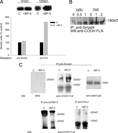
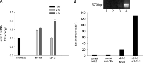
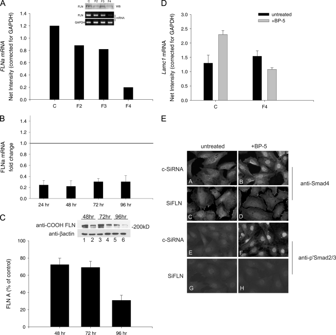
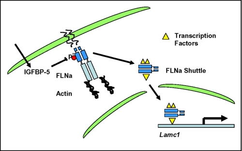
Insulin-like growth factor-binding protein-5 (IGFBP-5) has IGF-1-independent intranuclear effects that are poorly defined. Treatment of cells with IGFBP-5 induces migration, prevents apoptosis, and leads to increased laminin subunit transcription. Similarly, filamin A (FLNa), an actin-binding protein that participates in cell attachment, plays important additional roles in signal transduction and modulation of transcriptional responses. In this report, we show that IGFBP-5 leads to dephosphorylation of FLNa with subsequent FLNa cleavage. Following cleavage, there is enhanced recruitment of Smad3/4 to a C-terminal FLNa fragment with nuclear translocation and subsequent binding to the promoter region of the laminin gamma1 (lamc1) gene. FLNa knockdown prevents IGFBP-5-mediated increases in lamc1 transcription. These data indicate that IGFBP-5 induces formation of a FLNa-based nuclear shuttle that recruits transcription factors and regulates transcription of IGFBP-5 target genes. These studies provide new insights into the mechanisms whereby IGFBP-5 and FLNa exert intranuclear effects.


Figures

FIGURE 1.
FIGURE 1.
IGFBP-5 induces FLNa reorganization. A, MC were stained with Gt anti-FLNa before (panel A) and 4 (panel B), 8 (panel C), and 24 (panel D) h after the addition of IGFBP-5201–218. FLNa reorganizes, leaving the periphery of the cell and then concentrating in attachment sites, as the filopodial phenotype that is associated with IGFBP-5-induced migration develops. B, MC were stained for F-actin (phalloidin, red), FLNa (green), and nuclei (DAPPI, blue) before and at 2, 4, and 8 h after the addition of IGFBP-5201–218. As seen with the N-terminal anti-FLNa antibody, intact FLNa is diffusely present throughout the cytoplasm and co-localizes with F-actin (yellow) in a submembrane location at 2 h and in discrete focal adhesions as they develop after treatment with IGFBP-5. In contrast, staining with antibody specific for repeat 24 at the C terminus shows intense perinuclear staining followed by co-localization with DAPI, indicating an increase in nuclear staining. This pattern is less apparent by 8 h as the altered cellular phenotype develops. C-FLN, C-terminal FLNa; N-FLN, N-terminal FLNa.
FIGURE 2.
FIGURE 2.
Nuclear accumulation of C-FLNa. A, mesangial cells were examined by confocal microscopy following treatment with IGFBP-5 and staining with antibody specific for C-FLNa (repeats 16–24). Diffuse granular staining of the cytoplasm is seen in untreated cells (panel A). By 2 h after the addition of IGFBP-5201–218 (panel B) some cells show nuclear staining for FLNa and perinuclear clustering of FLNa (cut through center of cell). At 4 h after treatment, a cut through the bottom of the cells shows cytoplasmic staining (panel C), whereas the cut through the top of the cells shows predominantly nuclear staining (panel D). B, cells were transfected with empty vector (GFP-C) (panels A and B) or GFP-FLNa containing an insert for C-terminal repeats 16–24 (GFP-cFLNa) (panels C and D). The cells were untreated (-BP-5, panels A and C) or treated with IGFBP-5201–218 (+BP-5, panels B and D) for 4 h. IGFBP-5201–218 induced nuclear uptake of cFLNa but had no effect on the distribution of the control GFP-C.
FIGURE 3.
FIGURE 3.
IGFBP-5 induces fragmentation and nuclear translocation of FLNa. A, cytosolic (C) and nuclear (N) fractions were isolated from cells prior to and at 0.5, 1, 2, and 4 h after the addition of IGFBP-5201–218. Staining for FLNa (anti-COOH-FLNa) in Western blot shows the IGFBP5-induced loss of intact FLNa and appearance of fragments in the cytoplasm at 30 min and 1 h. Nuclear appearance of both intact FLNa and a 100-kDa fragment occur beginning at 1 h and are more pronounced at 2 and 4 h after the addition of IGFBP-5201–218. β-Actin (a cytosolic protein) and TFIIB (a nuclear protein) serve as loading controls and purification markers for the subcellular fractions. B, the nuclear fraction was isolated at 1 and 4 h after treatment with intact IGFBP-5 (I) or IGFBP-5201–218 (p) and subjected to Western blot using the anti-COOH-FLNa antibody. Both intact IGFBP-5 protein and peptide mediate nuclear uptake of a C-terminal fragment of FLNa. TFIIB serves as a loading control for the nuclear fraction.
FIGURE 4.
FIGURE 4.
IGFBP-5-induced FLNa dephosphorylation. Cytosolic fractions isolated at the times indicated following the addition of IGFBP5201–218 in control and cells pretreated with cyclosporine A were stained for intact FLNa and with antibodies to phospho-FLNa at serines 2113 (A) and 2152 (B). Bar graphs of the scanning densitometry results for p′FLN/total FLNa are shown. Although the total amount of FLNa detected did not significantly change within the first 30 min, the addition of IGFBP-5201–218 led to a significant reduction in the amount of p′FLNa detected at both serine residues. To examine the role of phosphatases in mediating the loss of p′FLNa, cells were treated with cyclosporine A (CsA) for 30 min prior to the addition of IGFBP-5201–218 and then stained for p′FLNa. Pretreatment with CsA prevented the dephosphorylation of FLNa at both Ser2113 (A) and Ser2152 (B). C, furthermore, the cells were stained with anti-FLNa before and at 2 and 4 h following the addition of IGFBP-5201–218. Pretreatment with CsA prevented the increase in nuclear staining that followed treatment with IGFBP-5201–218, which suggests that prevention of dephosphorylation prevents nuclear uptake of FLNa.
FIGURE 5.
FIGURE 5.
IGFBP-5201–218 induces increased binding of transcription factors to FLNa. A, lysates collected from control and IGFBP-5201–218-treated cells (after 4 h) were subjected to immunoprecipitation (IP) with either anti-Smad4 or anti-FLNa followed by Western blotting (WB) with anti-Smad4. The amount of Smad4 was equal; however, FLNa-bound Smad4 increased with IGFBP-5201–218 treatment. B, cytoplasmic (cyto) and nuclear (nuc) extracts were subjected to immunoprecipitation with anti-Smad4 at the times shown, followed by Western blotting for FLNa. The amount of Smad4 bound to FLNa increased within 30 min of treatment with IGFBP-5201–218, and an increase was detected in the nucleus by 1 h. C, immunoprecipitation of similar cell lysates showed that co-precipitation of FLNa with Smad3 and IGFBP-5 were also increased by treatment with IGFBP-5, whereas the content of FHL2 was unchanged.
FIGURE 6.
FIGURE 6.
Intact IGFBP-5 and IGFBP-5201–218-mediated increase in lamc1 mRNA is associated with FLN recruitment to the lamc1 promoter. A, effect of intact IGFBP-5 (BP-5 I) and IGFBP-5201–218 (BP-5p) on lamc1 mRNA. The results are expressed as fold change from the untreated control. The delayed and more robust response from intact IGFBP-5 suggests that cleavage may be required before it is taken up by the cell. B, ChIP assay shows that treatment with IGFBP-5201–218 induces recruitment of FLNa to the lamc1 gene. lamc1 DNA is not detected in precipitates from normal goat serum (NGtS) (lanes 2 and 4), nor in the absence of treatment with IGFBP-5 (lanes 1 and 2). The bar graph reflects net intensity of the reverse transcription-PCR products shown above.
FIGURE 7.
FIGURE 7.
siRNA-mediated knockdown of FLNa blocks IGFBP-5-induced lamc1 transcription and nuclear translocation of Smad. A, three separate FLNa siRNA sequences (F2, F3, and F4) were examined. Because F4 was the most effective, it was used in subsequent experiments. B, knockdown of FLNa mRNA with F4 persisted for 96 h (n = 3). C, the reduction in FLNa protein was maximal at 96 h (n = 3). D, in separate experiments using the time course defined above, FLNa knockdown with F4 blocks IGFBP-5201–218-induced increases in lamc1 mRNA as compared with the control siRNA (n = 3, p < 0.05, analysis of variance). E, as compared with cells transfected with a control siRNA (c-SiRNA), FLNa knockdown (SiFLN) was associated with loss of nuclear translocation of Smad4 and phospho-Smad2/3 that normally occurs following treatment with IGFBP-5.
FIGURE 8.
FIGURE 8.
Proposed schema. IGFBP-5 induces dephosphorylation of FLNa, rendering it susceptible to calpain cleavage. The C-terminal FLNa fragment recruits transcription factors and other transcriptional modulators, and the FLNa-based shuttle is translocated to the nucleus. Within the nucleus, the shuttle modulates transcription of IGFBP-5 target genes.

Similar articles

Cited by

References

    1. Mukherjee A., Wilson E. M., Rotwein P. (2008) Mol. Endocrinol. 22, 206–215 - PMC - PubMed
    1. Beattie J., Allan G. J., Lochrie J. D., Flint D. J. (2006) Biochem. J. 395, 1–19 - PMC - PubMed
    1. Schneider M. R., Wolf E., Hoeflich A., Lahm H. (2002) J. Endocrinol. 172, 423–440 - PubMed
    1. Parker A., Clarke J. B., Busby W. H., Jr., Clemmons D. R. (1996) J. Biol. Chem. 271, 13523–13529 - PubMed
    1. Booth B. A., Boes M., Andress D. L., Dake B. L., Kiefer M. C., Maack C., Linhardt R. J., Bar K., Caldwell E. E., Weiler J., Bar R. S. (1995) Growth Regul. 5, 1–17 - PubMed

Publication types

MeSH terms

LinkOut - more resources