

Parametric color coding of digital subtraction angiography
- PMID: 20167651
- PMCID: PMC7964185
- DOI: 10.3174/ajnr.A2020
Parametric color coding of digital subtraction angiography
Abstract
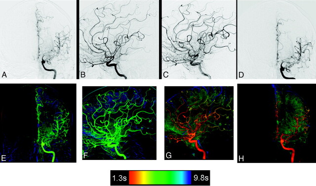
Background and purpose: Color has been shown to facilitate both visual search and recognition tasks. It was our purpose to examine the impact of a color-coding algorithm on the interpretation of 2D-DSA acquisitions by experienced and inexperienced observers.
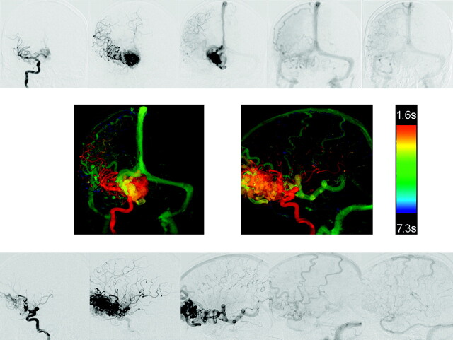
Materials and methods: Twenty-six 2D-DSA acquisitions obtained as part of routine clinical care from subjects with a variety of cerebrovascular disease processes were selected from an internal data base so as to include a variety of disease states (aneurysms, AVMs, fistulas, stenosis, occlusions, dissections, and tumors). Three experienced and 3 less experienced observers were each shown the acquisitions on a prerelease version of a commercially available double-monitor workstation (XWP, Siemens Healthcare). Acquisitions were presented first as a subtracted image series and then as a single composite color-coded image of the entire acquisition. Observers were then asked a series of questions designed to assess the value of the color-coded images for the following purposes: 1) to enhance their ability to make a diagnosis, 2) to have confidence in their diagnosis, 3) to plan a treatment, and 4) to judge the effect of a treatment. The results were analyzed by using 1-sample Wilcoxon tests.
Results: Color-coded images enhanced the ease of evaluating treatment success in >40% of cases (P < .0001). They also had a statistically significant impact on treatment planning, making planning easier in >20% of the cases (P = .0069). In >20% of the examples, color-coding made diagnosis and treatment planning easier for all readers (P < .0001). Color-coding also increased the confidence of diagnosis compared with the use of DSA alone (P = .056). The impact of this was greater for the naïve readers than for the expert readers.
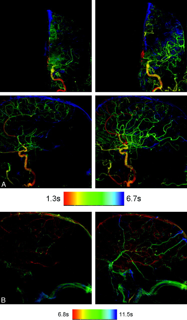
Conclusions: At no additional cost in x-ray dose or contrast medium, color-coding of DSA enhanced the conspicuity of findings on DSA images. It was particularly useful in situations in which there was a complex flow pattern and in evaluation of pre- and posttreatment acquisitions. Its full potential remains to be defined.
Figures
Comment in
-
Color-coded digital subtraction angiography: the end of a monochromatic era?AJNR Am J Neuroradiol. 2010 May;31(5):925-7. doi: 10.3174/ajnr.A2077. Epub 2010 Apr 15. AJNR Am J Neuroradiol. 2010. PMID: 20395396 Free PMC article. No abstract available.
References
-
- Cole BL, Maddocks JD, Sharpe K. Visual search and the conspicuity of coloured targets for colour vision normal and colour vision deficient observers. Clin Exp Optom 2004;87:294–304 - PubMed
-
- D'Zmura M. Color in visual search. Vision Res 1991;31:951–66 - PubMed
-
- Heintzen PH, Bürsch HJ, Hahne HJ, et al. Assessment of cardiovascular function by digital angiocardiography. J Am Coll Cardiol 1985:5:150S–57S - PubMed
-
- Cusma JT, Toggart EJ, Folts JD, et al. Digital subtraction angiograpic imaging of coronary flow reserve. Circulation 1987;75:461–72 - PubMed
-
- Bürsch HJ. Funktionsangiokardiographie. Kardiologie 1989;78(suppl 7):181–86 - PubMed
MeSH terms
LinkOut - more resources
Full Text Sources
Other Literature Sources
Medical