

Antitumor activity of an allosteric inhibitor of centromere-associated protein-E
- PMID: 20167803
- PMCID: PMC2851928
- DOI: 10.1073/pnas.0915068107
Antitumor activity of an allosteric inhibitor of centromere-associated protein-E
Abstract
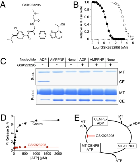
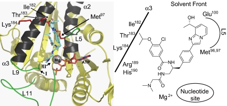
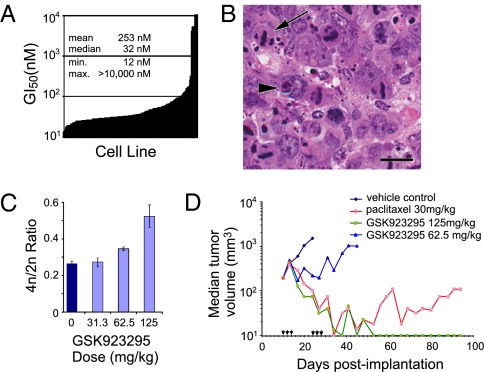
Centromere-associated protein-E (CENP-E) is a kinetochore-associated mitotic kinesin that is thought to function as the key receptor responsible for mitotic checkpoint signal transduction after interaction with spindle microtubules. We have identified GSK923295, an allosteric inhibitor of CENP-E kinesin motor ATPase activity, and mapped the inhibitor binding site to a region similar to that bound by loop-5 inhibitors of the kinesin KSP/Eg5. Unlike these KSP inhibitors, which block release of ADP and destabilize motor-microtubule interaction, GSK923295 inhibited release of inorganic phosphate and stabilized CENP-E motor domain interaction with microtubules. Inhibition of CENP-E motor activity in cultured cells and tumor xenografts caused failure of metaphase chromosome alignment and induced mitotic arrest, indicating that tight binding of CENP-E to microtubules is insufficient to satisfy the mitotic checkpoint. Consistent with genetic studies in mice suggesting that decreased CENP-E function can have a tumor-suppressive effect, inhibition of CENP-E induced tumor cell apoptosis and tumor regression.
Conflict of interest statement
Conflict of interest statement: All authors are current or former employees of Cytokinetics or GlaxoSmithKline and may hold stock in these companies.
Figures

Comment in
-
Targeting a kinetochore-associated motor protein to kill cancer cells.Proc Natl Acad Sci U S A. 2010 Mar 30;107(13):5699-700. doi: 10.1073/pnas.1001277107. Epub 2010 Mar 22. Proc Natl Acad Sci U S A. 2010. PMID: 20308538 Free PMC article. No abstract available.
Similar articles
-
Identification of benzo[d]pyrrolo[2,1-b]thiazole derivatives as CENP-E inhibitors.Biochem Biophys Res Commun. 2019 Nov 12;519(3):505-511. doi: 10.1016/j.bbrc.2019.09.028. Epub 2019 Sep 14. Biochem Biophys Res Commun. 2019. PMID: 31530389
-
Mitogen-activated protein kinase (MEK/ERK) inhibition sensitizes cancer cells to centromere-associated protein E inhibition.Int J Cancer. 2013 Feb 1;132(3):E149-57. doi: 10.1002/ijc.27781. Epub 2012 Sep 28. Int J Cancer. 2013. PMID: 22948716 Free PMC article.
-
Mitosis. Microtubule detyrosination guides chromosomes during mitosis.Science. 2015 May 15;348(6236):799-803. doi: 10.1126/science.aaa5175. Epub 2015 Apr 23. Science. 2015. PMID: 25908662 Free PMC article.
-
Mechanisms of kinesin-7 CENP-E in kinetochore-microtubule capture and chromosome alignment during cell division.Biol Cell. 2019 Jun;111(6):143-160. doi: 10.1111/boc.201800082. Epub 2019 Feb 26. Biol Cell. 2019. PMID: 30784092 Review.
-
Leaving no-one behind: how CENP-E facilitates chromosome alignment.Essays Biochem. 2020 Sep 4;64(2):313-324. doi: 10.1042/EBC20190073. Essays Biochem. 2020. PMID: 32347304 Free PMC article. Review.
Cited by
-
Microtubule-sliding modules based on kinesins EG5 and PRC1-dependent KIF4A drive human spindle elongation.Dev Cell. 2021 May 3;56(9):1253-1267.e10. doi: 10.1016/j.devcel.2021.04.005. Epub 2021 Apr 27. Dev Cell. 2021. PMID: 33910056 Free PMC article.
-
Mitosis-targeted anti-cancer therapies: where they stand.Cell Death Dis. 2012 Oct 18;3(10):e411. doi: 10.1038/cddis.2012.148. Cell Death Dis. 2012. PMID: 23076219 Free PMC article. Review.
-
Adaptive changes in the kinetochore architecture facilitate proper spindle assembly.Nat Cell Biol. 2015 Sep;17(9):1134-44. doi: 10.1038/ncb3223. Epub 2015 Aug 10. Nat Cell Biol. 2015. PMID: 26258631 Free PMC article.
-
Chromosome missegregation rate predicts whether aneuploidy will promote or suppress tumors.Proc Natl Acad Sci U S A. 2013 Oct 29;110(44):E4134-41. doi: 10.1073/pnas.1317042110. Epub 2013 Oct 16. Proc Natl Acad Sci U S A. 2013. PMID: 24133140 Free PMC article.
-
Small molecule screen for candidate antimalarials targeting Plasmodium Kinesin-5.J Biol Chem. 2014 Jun 6;289(23):16601-14. doi: 10.1074/jbc.M114.551408. Epub 2014 Apr 15. J Biol Chem. 2014. PMID: 24737313 Free PMC article.
References
-
- Wood KW, Chua P, Sutton D, Jackson JR. Centromere-associated protein E: A motor that puts the brakes on the mitotic checkpoint. Clin Cancer Res. 2008;14:7588–7592. - PubMed
-
- Yen TJ, Li G, Schaar BT, Szilak I, Cleveland DW. CENP-E is a putative kinetochore motor that accumulates just before mitosis. Nature. 1992;359:536–539. - PubMed
Publication types
MeSH terms
Substances
LinkOut - more resources
Full Text Sources
Other Literature Sources
