. 2010 Apr;176(4):1779-89.
doi: 10.2353/ajpath.2010.090738. Epub 2010 Feb 18.

Toll-like receptor 3 ligand dampens liver inflammation by stimulating Valpha 14 invariant natural killer T cells to negatively regulate gammadeltaT cells

Affiliations

Toll-like receptor 3 ligand dampens liver inflammation by stimulating Valpha 14 invariant natural killer T cells to negatively regulate gammadeltaT cells

Tommy R Gardner et al. Am J Pathol. 2010 Apr.

Abstract

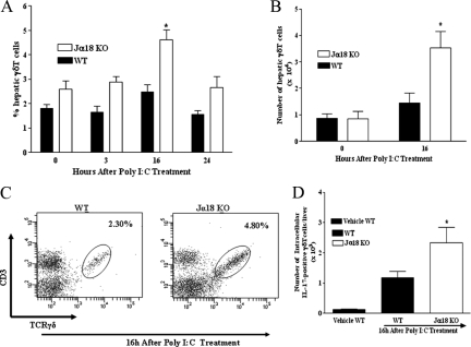
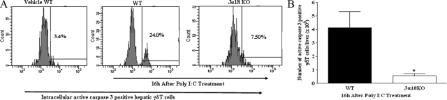
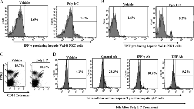
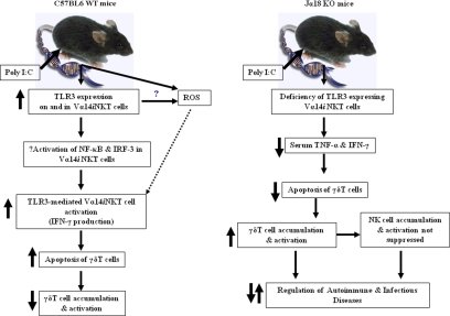
Valpha14 invariant natural killer T (Valpha14iNKT) cells are at the interface between the innate and adaptive immune responses and are thus critical for providing full engagement of host defense. We investigated the role of polyriboinosinic:polycytidylic acid (poly I:C), a replication-competent viral double-stranded RNA mimic and a specific agonist that recognizes the cellular sensor Toll-like receptor 3 (TLR3), in regulating Valpha14iNKT cell activation. We established for the first time that hepatic Valpha14iNKT cells up-regulate TLR3 extracellularly after poly I:C treatment. Notably, activation of TLR3-expressing hepatic Valpha14iNKT cells by a TLR3 ligand was suppressed by TLR3 deficiency. Our studies also revealed that Valpha14iNKT cell activation in response to poly I:C administration uniquely suppressed the accumulation and activation of intrahepatic gammadeltaT cells (but not natural killer cells) by inducing apoptosis. Furthermore, we established that activated hepatic Valpha14iNKT cells (via cytokines and possibly reactive oxygen species) influenced the frequency and absolute number of intrahepatic gammadeltaT cells, as evidenced by increased hepatic gammadeltaT cell accumulation in Valpha14iNKT cell-deficient mice after poly I:C treatment relative to wild-type mice. Thus, hepatic Valpha14iNKT cells and intrahepatic gammadeltaT cells are functionally linked on application of TLR3 agonist. Overall, our results demonstrate a novel and previously unrecognized anti-inflammatory role for activated hepatic Valpha14iNKT cells in negatively regulating intrahepatic gammadeltaT cell accumulation (probably through TLR3 signaling) and thereby preventing potentially harmful activation of intrahepatic gammadeltaT cells.


Figures

Figure 1
Figure 1
Vα14iNKT cell deficiency promotes hepatic γδT cell accumulation and activation after poly I:C treatment. C57BL/6 wild-type (WT) and Jα18 KO mice were treated with poly I:C at the indicated time points. Next, hepatic lymphoid cells were isolated from mice. Hepatic γδT cells were identified by FACS after cell surface staining with TCRγδ and CD3 mAbs (as described in Materials and Methods) and then gating on TCRγδ/CD3 double-positive T cells. The percentage (A), absolute number (B), and representative FACS dot plot (C) of hepatic γδT cells (ie, TCRγδ/CD3 double-positive T cells) are depicted. Results are presented as means ± SEM with four to six mice per group from two independent experiments. *P ≤ 0.05 versus all other groups. D: Absolute number of intracellular IL-17 expression by hepatic γδT cells was determined by FACS (as described in Materials and Methods). Data are shown as means ± SEM with three to six mice per group from two separate experiments. *P ≤ 0.05 versus vehicle-treated wild-type mice.
Figure 2
Figure 2
Activated hepatic Vα14iNKT cells promote apoptotic death of intrahepatic γδT cells after poly I:C treatment. Hepatic γδT cells were identified by FACS after cell surface staining with TCRγδ and CD3 mAbs. Hepatic γδT cells were fixed, permeabilized, and then stained intracellularly for active caspase 3. A representative FACS histogram (A) and absolute number (B) of intracellular active caspase 3-positive γδT cells are shown. Data are presented as means ± SEM with three to four mice per group from two independent experiments. *P ≤ 0.05 versus poly I:C-treated wild-type (WT) mice.
Figure 3
Figure 3
Contribution of Vα14iNKT cell-derived cytokines to apoptosis of intrahepatic γδT cells after poly I:C treatment. A and B: C57BL/6 wild-type mice were treated with poly I:C or vehicle for 16 hours, and isolated hepatic Vα14iNKT cells were identified by FACS after cell surface staining with TCRβ mAb and PBS57-CD1d tetramer (described in Materials and Methods). The cells were fixed, permeabilized, and then stained intracellularly for IFN-γ or TNF. Representative FACS histograms demonstrating IFN-γ-positive hepatic Vα14iNKT cells and TNF-positive hepatic Vα14iNKT cells are shown in A and B, respectively, from two independent experiments of four mice per group. C: FACS dot plot (from two independent experiments of three mice per group) depicts hepatic Vα14iNKT cell profiles from poly I:C or vehicle-treated C57BL/6 wild-type mice at the 16-hour time point. D: C57BL/6 wild-type mice were pretreated with murine IFN-γ antiserum, murine TNF mAb, or control antibody before poly I:C treatment, and 16 hours later isolated hepatic γδT cells were fixed, permeabilized, and then stained intracellularly for active caspase 3. A representative FACS histogram highlighting active caspase 3-positive hepatic γδT cells from two independent experiments of four mice per group is shown.
Figure 4
Figure 4
Role of endogenous ROS in hepatic Vα14iNKT cell activation on poly I:C treatment. The ROS scavenger, NAC (300 mg/kg i.p.), was administered simultaneously with poly I:C in C57BL/6 wild-type (WT) mice or Jα18 KO mice. Control mice received vehicle PBS. A: Hepatic Vα14iNKT cells were identified by FACS using the CD1d tetramer and TCRβ mAb (see Materials and Methods) 16 hours after poly I:C treatment and then were stained for intracellular IFN-γ (as a marker for Vα14iNKT cell activation) after cell permeabilization. Results are presented as means ± SEM with four mice per group from two separate experiments. *P ≤ 0.05 versus vehicle. B: C57BL/6 wild-type mice and Jα18 KO mice were treated concurrently with NAC/poly I:C or PBS/poly I:C for intrahepatic γδT cell accumulation assessment by FACS 16 hours later. Data are presented as means ± SEM with four to six mice per group from two independent studies. ***P ≤ 0.05 versus all other groups; **P ≤ 0.05 versus vehicle-treated wild-type mice (no poly I:C) and poly I:C/PBS treated wild-type mice; *P ≤ 0.05 versus vehicle-treated wild-type mice (no poly I:C) and poly I:C/PBS-treated wild-type mice.
Figure 5
Figure 5
Hepatic Vα14iNKT cell TLR3 expression before and after poly I:C treatment. B6129SF2/J wild-type mice were treated with poly I:C or vehicle for 16 hours and extracellular and intracellular TLR3 expression by hepatic Vα14iNKT cells was evaluated by flow cytometry as described in Materials and Methods. Representative FACS histograms are shown from two independent experiments.
Figure 6
Figure 6
Effects of TLR3 deficiency on hepatic Vα14iNKT cell cytokine production in response to poly I:C application. TLR3-deficient mice and B6129SF2/J wild-type (WT) mice were treated with poly I:C for 16 hours for the isolation and identification of hepatic Vα14iNKT cells by FACS. Intracellular staining was performed on hepatic Vα14iNKT cells (after cell permeabilization) using fluorochrome-labeled cytokine antibodies (see Materials and Methods) to determine the percentage and absolute numbers of both IFN-γ expressing Vα14iNKT cells (A and B) as well as TNF-producing Vα14iNKT cells (C and D). The dotted line in C and D represents the value in wild-type mice treated with vehicle. All results are presented as means ± SEM with three to four mice per group from two independent experiments. *P ≤ 0.05 versus wild-type mice.
Figure 7
Figure 7
A schematic model to illustrate the mechanisms of TLR3 ligand-dependent hepatic Vα14iNKT cell activation and the subsequent induction of apoptotic death of intrahepatic γδT cells on poly I:C application. Left panel: Vα14iNKT cells up-regulate TLR3 extracellularly on C57BL/6 wild-type (WT) mice on poly I:C treatment and activation of TLR3-expressing Vα14iNKT cells by poly I:C-induced Vα14iNKT cell cytokine production is through a signaling pathway, which could be dependent on the transcription factors nuclear factor-κB (NF-κB) and interferon regulatory factor-3 (IRF-3). Notably, these responses were suppressed by TLR3 deficiency. In addition, our study also revealed that the functional consequences of hepatic Vα14iNKT cell activation by the TLR3 ligand, poly I:C, is the negative regulation of γδT cells as shown by decreased accumulation of activated intrahepatic γδT cells due to apoptosis by Vα14iNKT cell-derived mediators (ie, cytokines and possibly ROS). Right panel: In contrast with that of C57BL/6 wild-type mice, poly I:C treatment of Jα18 KO mice was associated with lower serum levels of prototypical Vα14iNKT cell-derived cytokines (ie, TNF and IFN-γ), most likely due to a deficiency of TLR3-expressing activated hepatic Vα14iNKT cells. The functional consequence of lower cytokine levels is the suppression of apoptotic death of intrahepatic γδT cells (and thus increased survival of intrahepatic γδT cells) as shown by increased accumulation of γδT cells in the livers of poly I:C-treated Jα18 KO mice. Notably, the lack of suppression of hepatic NK cell accumulation and activation in Jα18 KO mice after poly I:C treatment may be due in part to the increased accumulation of γδT cells in the livers of these mice because we recently demonstrated that γδT cells promote NK cell accumulation in the liver after poly I:C treatment. Our study provides the first direct evidence that intrahepatic Vα14iNKT cells and γδT cells are functionally linked via TLR3 signaling on application of poly I:C because activated intrahepatic Vα14iNKT cells negatively regulate the recruitment, activation, and potentially harmful effector function(s) of intrahepatic γδT cells on application of the TLR3 ligand, poly I:C.

Similar articles

Cited by

References

    1. Van Kaer L. α-Galactosylceramide therapy for autoimmune diseases: prospects and obstacles. Nat Rev Immunol. 2005;5:31–42. - PubMed
    1. Kronenberg M. Toward an understanding of NKT cell biology: progress and paradoxes. Annu Rev Immunol. 2005;23:877–900. - PubMed
    1. Wilson MT, Singh AK, Van Kaer L. Immunotherapy with ligands of natural killer T cells. Trends Mol Med. 2002;8:225–231. - PubMed
    1. Wilson MT, Van Kaer L. Natural killer T cells as targets for therapeutic intervention in autoimmune diseases. Curr Pharm Des. 2003;9:201–220. - PubMed
    1. Bendelac A, Savage PB, Teyton L. The biology of NKT cells. Annu Rev Immunol. 2007;25:297–336. - PubMed

Publication types