Review
. 2010 Feb 19;106(3):463-78.
doi: 10.1161/CIRCRESAHA.109.208801.

Sent to destroy: the ubiquitin proteasome system regulates cell signaling and protein quality control in cardiovascular development and disease

Affiliations
Review

Sent to destroy: the ubiquitin proteasome system regulates cell signaling and protein quality control in cardiovascular development and disease

Monte S Willis et al. Circ Res. .

Abstract

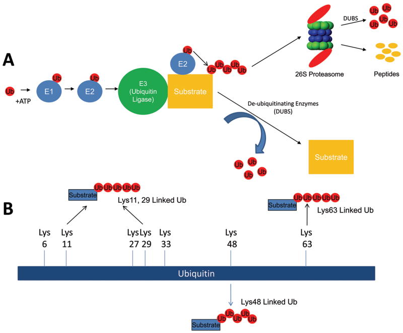
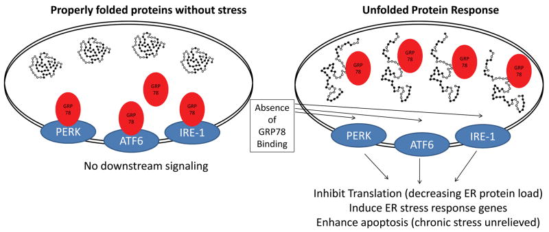
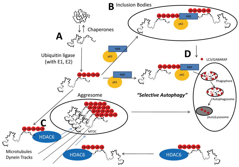
The ubiquitin proteasome system (UPS) plays a crucial role in biological processes integral to the development of the cardiovascular system and cardiovascular diseases. The UPS prototypically recognizes specific protein substrates and places polyubiquitin chains on them for subsequent destruction by the proteasome. This system is in place to degrade not only misfolded and damaged proteins, but is essential also in regulating a host of cell signaling pathways involved in proliferation, adaptation to stress, regulation of cell size, and cell death. During the development of the cardiovascular system, the UPS regulates cell signaling by modifying transcription factors, receptors, and structural proteins. Later, in the event of cardiovascular diseases as diverse as atherosclerosis, cardiac hypertrophy, and ischemia/reperfusion injury, ubiquitin ligases and the proteasome are implicated in protecting and exacerbating clinical outcomes. However, when misfolded and damaged proteins are ubiquitinated by the UPS, their destruction by the proteasome is not always possible because of their aggregated confirmations. Recent studies have discovered how these ubiquitinated misfolded proteins can be destroyed by alternative "specific" mechanisms. The cytosolic receptors p62, NBR, and histone deacetylase 6 recognize aggregated ubiquitinated proteins and target them for autophagy in the process of "selective autophagy." Even the ubiquitination of multiple proteins within whole organelles that drive the more general macro-autophagy may be due, in part, to similar ubiquitin-driven mechanisms. In summary, the crosstalk between the UPS and autophagy highlight the pivotal and diverse roles the UPS plays in maintaining protein quality control and regulating cardiovascular development and disease.


Figures

Figure 1
Figure 1. The ubiquitin proteasome system at a glance
The ubiquitin proteasome is a system of enzymes that places ubiquitin (chains) on specific protein substrates to target them for degradation, change their localization, and/or enhance their activity. (A) Free monoubiquitin is activated by the E1 enzyme in an ATP-dependent manner and transferred to the E2 enzyme. The specificity of the system is in the E3 (ubiquitin ligase) that mediates the transfer of one or more ubiquitin moieties sequentially to form ubiquitin chains on the substrate. The canonical lysine chains linked by their lysine48 (Lys48) are recognized by the 26S proteasome which degrades the protein into constituent peptides and free ubiquitin. (B) The role of noncanonical polyubiquitination (i.e. Lys63-linked ubiquitin chains) is increasingly being described in the cardiovascular system as a way to regulate protein (i.e. transcription factors) activity. A total of seven lysine moieties exist in ubiquitin which can be used for chain formation (Lys6, Lys11, Lys27, Lys29, Lys 33, Lys48, Lys63). A number of studies discussed in this review reveal a role for more linear Lys63 chains, in addition to canonical Lys48 linkages in the cardiovascular system. However, the significance of Lys6, Lys11, Lys 27, Lys29, Lys33 and branching/complex ubiquitin chains has not been elucidated in general.
Figure 2
Figure 2. Endoplasmic reticulum stress and the unfolded protein response
In response to stress, proteins synthesized in the rough endoplasmic reticulum are refolded by resident molecular chaperones GP78 (glucose-regulated protein-78). Upon refolding, GRP78 looses its association with the luminal domains of the PKR-like ER kinase (PERK), activating transcription factor-6 (ATF6), and inositol-required enzyme-1 (IRE-1). This leads to PERK, ATF6, and IRE-1 activation and downstream activation of ER stress response genes which help stabilize the misfolding of proteins, including ER-targeted chaperones. If the unfolded proteins are not adequately removed after the activation of the UPR, signaling pathways for apoptosis can be activated. Adapted from: Glembotski et al., 2007 and Glembotski et al,. 2008.
Figure 3
Figure 3. Selective Autophagy through recognition of misfolded and ubiquitinated proteins through NRB1 and p62
(A) Stress-induced misfolding of proteins is a constant threat to the well-being of the cell. Chaperones continually refold proteins by recognizing the hydrophobic regions of the protein exposed during stress. Heat shock protein-ubiquitin ligase complexes promote folding, but if this is not possible they enhance the ubiquitination of recognized substances effectively targeting proteins for either proteasome degradation (see Figure 1) or by selective autophagy. (B) Misfolded ubiquitinated proteins can polymerize to form inclusion bodies and (C) aggresomes which form from the transport of aggregated ubiquitinated proteins that are transported via dynein on microtubule tracks. (D) “Selective autophagy” is the catabolism of macromolecule and organelles based on the recognition of ubiquitination chains on proteins which plays an important role in maintaining protein quality control in the cell. MTOC, Microtubule organizing centers. Adapted from: Kirkin, et al., 2009 and Larmark et al., 2009 .

References

    1. Pickart CM, Fushman D. Polyubiquitin chains: polymeric protein signals. Curr Opin Chem Biol. 2004;8(6):610–616. - PubMed
    1. Li HH, Willis MS, Lockyer P, Miller N, McDonough H, Glass DJ, Patterson C. Atrogin-1 inhibits Akt-dependent cardiac hypertrophy in mice via ubiquitin-dependent coactivation of Forkhead proteins. J Clin Invest. 2007;117(11):3211–3223. - PMC - PubMed
    1. Spence J, Sadis S, Haas AL, Finley D. A ubiquitin mutant with specific defects in DNA repair and multiubiquitination. Mol Cell Biol. 1995;15(3):1265–1273. - PMC - PubMed
    1. Haglund K, Shimokawa N, Szymkiewicz I, Dikic I. Cbl-directed monoubiquitination of CIN85 is involved in regulation of ligand-induced degradation of EGF receptors. Proc Natl Acad Sci U S A. 2002;99(19):12191–12196. - PMC - PubMed
    1. Leithe E, Rivedal E. Ubiquitination and down-regulation of gap junction protein connexin-43 in response to 12-O-tetradecanoylphorbol 13-acetate treatment. J Biol Chem. 2004;279(48):50089–50096. - PubMed

Publication types

MeSH terms