Liver membrane proteome glycosylation changes in mice bearing an extra-hepatic tumor
- PMID: 20167946
- PMCID: PMC3186205
- DOI: 10.1074/mcp.M900538-MCP200
Liver membrane proteome glycosylation changes in mice bearing an extra-hepatic tumor
Abstract
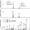
Cancer is well known to be associated with alterations in membrane protein glycosylation (Bird, N. C., Mangnall, D., and Majeed, A. W. (2006) Biology of colorectal liver metastases: A review. J. Surg. Oncol. 94, 68-80; Dimitroff, C. J., Pera, P., Dall'Olio, F., Matta, K. L., Chandrasekaran, E. V., Lau, J. T., and Bernacki, R. J. (1999) Cell surface n-acetylneuraminic acid alpha2,3-galactoside-dependent intercellular adhesion of human colon cancer cells. Biochem. Biophys. Res. Commun. 256, 631-636; and Arcinas, A., Yen, T. Y., Kebebew, E., and Macher, B. A. (2009) Cell surface and secreted protein profiles of human thyroid cancer cell lines reveal distinct glycoprotein patterns. J. Proteome Res. 8, 3958-3968). Equally, it has been well established that tumor-associated inflammation through the release of pro-inflammatory cytokines is a common cause of reduced hepatic drug metabolism and increased toxicity in advanced cancer patients being treated with cytotoxic chemotherapies. However, little is known about the impact of bearing a tumor (and downstream effects like inflammation) on liver membrane protein glycosylation. In this study, proteomic and glycomic analyses were used in combination to determine whether liver membrane protein glycosylation was affected in mice bearing the Engelbreth-Holm Swarm sarcoma. Peptide IPG-IEF and label-free quantitation determined that many enzymes involved in the protein glycosylation pathway specifically; mannosidases (Man1a-I, Man1b-I and Man2a-I), mannoside N-acetylglucosaminyltransferases (Mgat-I and Mgat-II), galactosyltransferases (B3GalT-VII, B4GalT-I, B4GalT-III, C1GalT-I, C1GalT-II, and GalNT-I), and sialyltransferases (ST3Gal-I, ST6Gal-I, and ST6GalNAc-VI) were up-regulated in all livers of tumor-bearing mice (n = 3) compared with nontumor bearing controls (n = 3). In addition, many cell surface lectins: Sialoadhesin-1 (Siglec-1), C-type lectin family 4f (Kupffer cell receptor), and Galactose-binding lectin 9 (Galectin-9) were determined to be up-regulated in the liver of tumor-bearing compared with control mice. Global glycan analysis identified seven N-glycans and two O-glycans that had changed on the liver membrane proteins derived from tumor-bearing mice. Interestingly, α (2,3) sialic acid was found to be up-regulated on the liver membrane of tumor-bearing mice, which reflected the increased expression of its associated sialyltransferase and lectin receptor (siglec-1). The overall increased sialylation on the liver membrane of Engelbreth-Holm Swarm bearing mice correlates with the increased expression of their associated glycosyltransferases and suggests that glycosylation of proteins in the liver plays a role in tumor-induced liver inflammation.
Figures


Similar articles
-
Increased alpha2,6 sialylation of N-glycans in a transgenic mouse model of hepatocellular carcinoma.Cancer Res. 1997 Oct 1;57(19):4249-56. Cancer Res. 1997. PMID: 9331085
-
Tumour suppressor p16(INK4a) - anoikis-favouring decrease in N/O-glycan/cell surface sialylation by down-regulation of enzymes in sialic acid biosynthesis in tandem in a pancreatic carcinoma model.FEBS J. 2012 Nov;279(21):4062-80. doi: 10.1111/febs.12001. FEBS J. 2012. PMID: 22943525
-
Terminal Galactosylation and Sialylation Switching on Membrane Glycoproteins upon TNF-Alpha-Induced Insulin Resistance in Adipocytes.Mol Cell Proteomics. 2016 Jan;15(1):141-53. doi: 10.1074/mcp.M115.054221. Epub 2015 Nov 4. Mol Cell Proteomics. 2016. PMID: 26537798 Free PMC article.
-
Significance of β-Galactoside α2,6 Sialyltranferase 1 in Cancers.Molecules. 2015 Apr 24;20(5):7509-27. doi: 10.3390/molecules20057509. Molecules. 2015. PMID: 25919275 Free PMC article. Review.
-
Galectin-Binding O-Glycosylations as Regulators of Malignancy.Cancer Res. 2015 Aug 15;75(16):3195-202. doi: 10.1158/0008-5472.CAN-15-0834. Epub 2015 Jul 29. Cancer Res. 2015. PMID: 26224120 Free PMC article. Review.
Cited by
-
Porcine sialoadhesin: a newly identified xenogeneic innate immune receptor.Am J Transplant. 2012 Dec;12(12):3272-82. doi: 10.1111/j.1600-6143.2012.04247.x. Epub 2012 Sep 7. Am J Transplant. 2012. PMID: 22958948 Free PMC article.
-
Deciphering the Properties and Functions of Glycoproteins Using Quantitative Proteomics.J Proteome Res. 2023 Jun 2;22(6):1571-1588. doi: 10.1021/acs.jproteome.3c00015. Epub 2023 Apr 3. J Proteome Res. 2023. PMID: 37010087 Free PMC article. Review.
-
The Influence of Clusterin Glycosylation Variability on Selected Pathophysiological Processes in the Human Body.Oxid Med Cell Longev. 2022 Aug 28;2022:7657876. doi: 10.1155/2022/7657876. eCollection 2022. Oxid Med Cell Longev. 2022. PMID: 36071866 Free PMC article. Review.
-
Glycomics and glycoproteomics of membrane proteins and cell-surface receptors: Present trends and future opportunities.Electrophoresis. 2016 Jun;37(11):1407-19. doi: 10.1002/elps.201500552. Epub 2016 Mar 29. Electrophoresis. 2016. PMID: 26872045 Free PMC article. Review.
-
DNA methylome and transcriptome alterations and cancer prevention by curcumin in colitis-accelerated colon cancer in mice.Carcinogenesis. 2018 May 3;39(5):669-680. doi: 10.1093/carcin/bgy043. Carcinogenesis. 2018. PMID: 29547900 Free PMC article.
References
-
- Bird N. C., Mangnall D., Majeed A. W. (2006) Biology of colorectal liver metastases: A review. J. Surg. Oncol. 94, 68–80 - PubMed
-
- Dimitroff C. J., Pera P., Dall'Olio F., Matta K. L., Chandrasekaran E. V., Lau J. T., Bernacki R. J. (1999) Cell surface n-acetylneuraminic acid alpha2,3-galactoside-dependent intercellular adhesion of human colon cancer cells. Biochem. Biophys. Res. Commun. 256, 631–636 - PubMed
-
- Washburn M. P., Wolters D., Yates J. R., 3rd (2001) Large-scale analysis of the yeast proteome by multidimensional protein identification technology. Nat. Biotechnol. 19, 242–247 - PubMed
-
- Wang W., Guo T., Rudnick P. A., Song T., Li J., Zhuang Z., Zheng W., Devoe D. L., Lee C. S., Balgley B. M. (2007) Membrane proteome analysis of microdissected ovarian tumor tissues using capillary isoelectric focusing/reversed-phase liquid chromatography-tandem MS. Anal. Chem. 79, 1002–1009 - PubMed
Publication types
MeSH terms
Substances
LinkOut - more resources
Full Text Sources
Research Materials

