. 2010 Jan-Feb;2(1):53-66.
doi: 10.4161/mabs.2.1.10788. Epub 2010 Jan 27.

Neutralizing epitopes of the SARS-CoV S-protein cluster independent of repertoire, antigen structure or mAb technology

Affiliations

Neutralizing epitopes of the SARS-CoV S-protein cluster independent of repertoire, antigen structure or mAb technology

Jody D Berry et al. MAbs. 2010 Jan-Feb.

Abstract

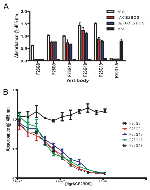
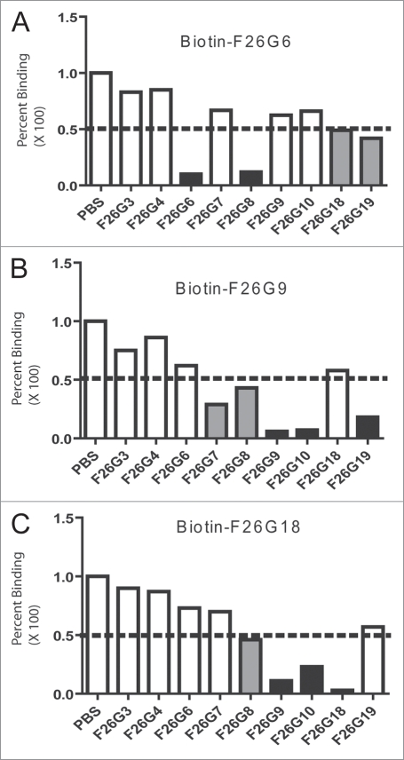
Neutralizing antibody responses to the surface glycoproteins of enveloped viruses play an important role in immunity. Many of these glycoproteins, including the severe acute respiratory syndrome-coronavirus (SARS-CoV) spike (S) protein form trimeric units in the membrane of the native virion. There is substantial experimental and pre-clinical evidence showing that the S protein is a promising lead for vaccines and therapeutics. Previously we generated a panel of monoclonal antibodies (mAbs) to whole inactivated SARS-CoV which neutralize the virus in vitro. Here, we define their specificity and affinity, map several of their epitopes and lastly characterise chimeric versions of them. Our data show that the neutralizing mAbs bind to the angiotensin-converting enzyme 2 (ACE2) receptor-binding domain (RBD) of the SARS S protein. Three of the chimeric mAbs retain their binding specificity while one conformational mAb, F26G19, lost its ability to bind the S protein despite high level expression. The affinity for recombinant S is maintained in all of the functional chimeric versions of the parental mAbs. Both parental mAb F26G18 and the chimeric version neutralize the TO R2 strain of SARS-CoV with essentially identical titres (2.07 and 2.47 nM, respectively). Lastly, a comparison with other neutralizing mAbs to SARS-CoV clearly shows that the dominance of a 33 amino acid residue loop of the SARS-CoV RBD is independent of repertoire, species, quaternary structure, and importantly, the technology used to derive the mAbs. In cases like this, the dominance of a compact RBD antigenic domain and the central role of the S protein in pathogenesis may inherently create immunoselection pressure on viruses to evolve more complex evasion strategies or die out of a host species. The apparent simplicity of the mechanism of SARS-CoV neutralization is in stark contrast to the complexity shown by other enveloped viruses.


Figures

Figure 1
Figure 1
ELISA Epitope characterisation of murine F26 series mAbs. All the F26 series mAbs tested bind to rFS and F26G8 is the only mAb that does not bind to the ACE2BD region, as demonstrated by lack of binding to either rACE2BDS or dgrACE2BDS (A). These results were confirmed by a competitive with rFS coating and dgrACE2BDS inhibition which clearly shows a reduction in signal with increasing dgrACE2BDS protein concentration for all mAbs except F26G8 (B).
Figure 2
Figure 2
Competition ELISA with biotinylated mAbs to whole inactivated SARs-CoV. The biotinylated mAbs F26G6 (A), F26G9 (B) and F26G18 (C), were evaluated for the ability to bind in the presence of competing mAbs. signal was detected using goat anti-mouse Fc gamma hrp and normalized to 100% binding in the presence of PBS. White bars, less than 0–50% inhibition; Gray bars, around 50% inhibition of binding; Black bars, >75% inhibition of the biotinylated mAb.
Figure 3
Figure 3
ELISA epitope characterisation of chimeric mAbs. Direct ELISA (A) shows that all the chimeric mAbs bind strongly to rFS, while rACE2BDS and rdgACE2BDS bind with varying degrees. Competitive ELISA (B) shows that rdgACE2BDS successfully competes with rFS for antibody binding for all the chimerics. B-inset shows western immunoblotting on rFS (2), rACE2BDS (3) and rPA (4) using chimeric antibody 18H18L. The chimeric antibody binds to both rFS and rACE2BDS, without binding rPA. Lane 1 is the molecular weight marker, with the values in kiloDaltons (kDa) on the left hand side.
Figure 4
Figure 4
Epitope Mapping of murine mAbs F26G8 and F26G18. (A) Pepscan Mapping of murine mAbs F26G8 and F26G18 on overlapping pin peptides spanning the S1 region (A) inset, Random Phage peptide mapping of mAb F26G8. Mimotope identification of anti-SARS monoclonal antibody, F26G8 after the second selection from Ph.D-12-mer phage displayed random peptide library mapping. Above the clone sequences is shown the actual S protein motif sequence. (B) inset, Direct ELISA on tethered peptide in an ELISA and (B), a competitive soluble peptide ELISA on rFS which confirms that mAbs F26G8 and F26G18 bind S1 with critical contacts in regions aa612–620 and aa460–476, respectively.
Figure 5
Figure 5
Peptide Mapping of 18H18L. Pin Peptide Mapping (A) and direct peptide binding ELISA (B) results are inconclusive compared to background (black bar, G18PEP; white bar, G18PEPScr; grey bar, G8PEP; hatched bar, G8PEPScr), while the competitive ELISA (C) demonstrates that 18H18L binds the aa460–476 peptide.
Figure 6
Figure 6
Depiction of the SARS-CoV Achilles heel. A schematic depicting the location of neutralizing epitopes and binding domains on the 193 amino acid receptor binding domain (RBD) of the S1 protein (Li et al. 2005). (A) Contact residues of ACE2 and mAbs which interrupt S1 binding to ACE2 are depicted with blue filled boxes. The DC-SIGN and mAbs which are known to block S1 binding to DC-SIGN are depicted with a pink filled box. The mAbs F26G18 and F26G19 were raised in immune response to native SARS-CoV spike protein (whole virus) and are outlined in red lined boxes. mAbs with linear epitopes are shown with solid lined boxes. mAbs with conformation epitopes have dashed lines on their boxes to indicate contact residues fall in the region as determined from co-crystal structure with S1 proteins. (B) The core 33 amino acid residues containing the known critical contacts of neutralizing mAbs and the ACE-2 receptor in the RBD (green dashed box). This immunodominant determinant corresponds to amino acid residues 460–492 of the S1 protein (green dashed box). This region is shown to illustrate the diversity of recognition of this compact region as well as the proximity of the contact residues of neutralizing mAbs in relation to the contact residues of the ACE2 host cellular receptor. Occlusion of the receptor is the main mechanisms of SARS virus neutralization for mAbs binding in this region. The boxes show the minimal epitope contact “footprint” in this region either from crystal structures and or other epitope mapping strategies for each of: ACE2 (red broken box); conformational mAb 80R (blue dotted box); conformational mAb F26G19 (black dashed box, 486–492); conformational mAb m396 main contact residues (brown box); and the linear epitope of mAb F26G18 (black box, 460–476). The known contact residues for ACE-2 are color coded red. Data has been extracted from this paper and other previously published articles.,,,,,

Similar articles

Cited by

References

    1. Berry JD, Jones S, Drebot MA, Andonov A, Sabara M, Yuan X, et al. Development and characterisation of neutralising monoclonal antibody to the SARS-coronavirus. J Virol Methods. 2004;120:87–96. - PMC - PubMed
    1. Gubbins MJ, Plummer FA, Yuan XY, Johnstone D, Drebot M, Andonova M, et al. Molecular characterization of a panel of murine monoclonal antibodies specific for the SARS-coronavirus. Mol Immunol. 2005;42:125–136. - PMC - PubMed
    1. Stadler K, Masignani V, Eickmann M, Becker S, Abrignani S, Klenk HD, et al. SARS—beginning to understand a new virus. Nat Rev Microbiol. 2003;1:209–218. - PMC - PubMed
    1. Li F, Li W, Farzan M, Harrison SC. Structure of SARS coronavirus spike receptor-binding domain complexed with receptor. Science. 2005;309:1864–1868. - PubMed
    1. Chakraborti S, Prabakaran P, Xiao X, Dimitrov DS. The SARS coronavirus S glycoprotein receptor binding domain: fine mapping and functional characterization. Virol J. 2005;2:73. - PMC - PubMed

Publication types

MeSH terms

LinkOut - more resources