. 2010 Jun;30(6):958-65.
doi: 10.1097/IAE.0b013e3181c9696a.

Accuracy of retinopathy of prematurity diagnosis by retinal fellows

Affiliations

Accuracy of retinopathy of prematurity diagnosis by retinal fellows

R V Paul Chan et al. Retina. 2010 Jun.

Abstract

Purpose: The purpose of this study was to measure the accuracy of retinopathy of prematurity (ROP) diagnosis by retinal fellows.

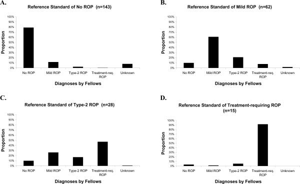
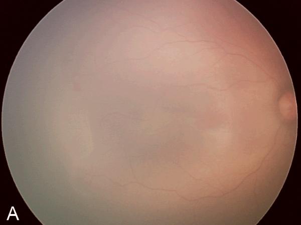
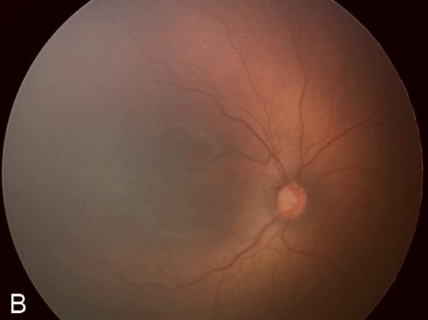
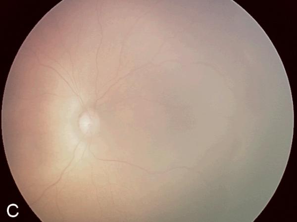
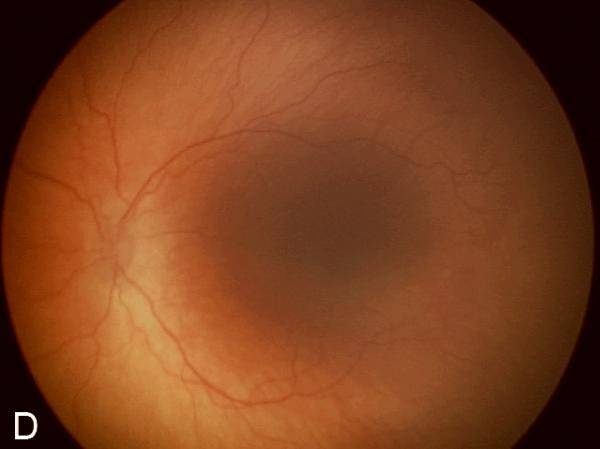
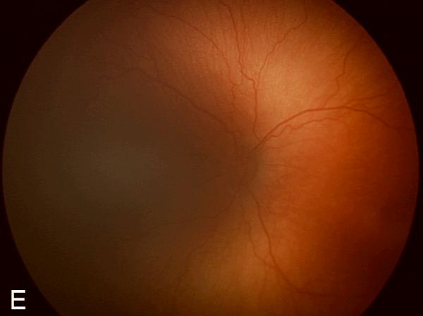
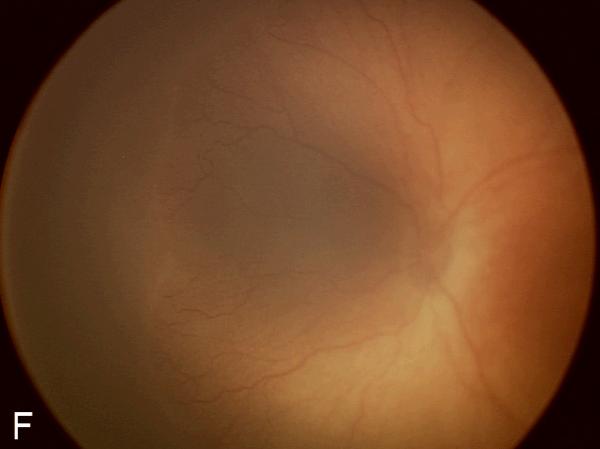
Methods: An atlas of 804 retinal images was captured from 248 eyes of 67 premature infants with a wide-angle camera (RetCam-II, Clarity Medical Systems, Pleasanton, CA). Images were uploaded to a study Web site, from which an expert pediatric retinal specialist and 7 retinal fellows independently provided a diagnosis (no ROP, mild ROP, type 2 ROP, or treatment-requiring ROP) for each eye. The sensitivity and specificity of each retinal fellow were calculated and subsequently compared with a reference standard of diagnosis by an expert pediatric retinal specialist.

Results: For detection of type 2 or worse ROP by fellows, mean (range) sensitivity was 0.751 (0.512-0.953), and specificity was 0.841 (0.707-0.976). For detection of treatment-requiring ROP, mean (range) sensitivity was 0.914 (0.667-1.000), and specificity was 0.871 (0.678-0.987).

Conclusion: In general, fellows showed high accuracy for detecting ROP. However, 3 of 7 fellows achieved <80% sensitivity for diagnosis of type 2 or worse ROP, and 2 of 7 achieved <90% sensitivity for diagnosis of treatment-requiring ROP. This could lead to undermanagement and undertreatment of clinically significant disease and raises potential concerns about the quality of ROP screening examinations performed by less-experienced examiners.


Figures

Figure 1
Figure 1. Mean distribution of ROP diagnoses by 7 retinal fellows
(A) Among the 143 eyes with a reference standard diagnosis of no ROP, the 7 fellows diagnosed no ROP in 113 (79%) eyes. (B) Among the 62 eyes with a reference standard diagnosis of mild ROP, the 7 fellows diagnosed mild ROP in 38 (61%) eyes. (C) Among the 28 eyes with a reference standard diagnosis of type-2 prethreshold ROP, the 7 fellows diagnosed type-2 prethreshold ROP in 5 (17%) eyes. (D) Among the 15 eyes with a reference standard diagnosis of treatment-requiring ROP, the 7 fellows diagnosed treatment-requiring ROP in 14 (91%) eyes.
Figure 2
Figure 2. Examples of study images that were frequently misdiagnosed by retinal fellows
(A), (B), and (C) display temporal, posterior, and nasal images from an infant diagnosed as mild ROP by reference standard exam diagnosis and was diagnosed as type-2 ROP by 5/7 (71%) fellows, and as treatment-requiring ROP by 2/7 (29%) fellows. (D), (E), and (F) display temporal, posterior, and nasal images from an infant diagnosed as type-2 ROP by reference standard exam diagnosis and was diagnosed as no ROP by 2/7 (29%) fellows, as type-2 ROP by 1/7 (14%) fellows, and as treatment-requiring ROP by 4/7 (57%) fellows.

Similar articles

Cited by

References

    1. Flynn JT, Bancalari E, Bachynski BN, et al. Retinopathy of prematurity. Diagnosis, severity, and natural history. Ophthalmology. 1987;94:620–629. - PubMed
    1. Martin JA, Hamilton BE, Sutton PD, et al. Births: final data for 2005. Natl Vital Stat Rep. 2007;56:1–103. - PubMed
    1. National Eye Institute Retinopathy of Prematurity. [Accessed May 16, 2008]. Available at: http://www.nei.nih.gov/health/rop/.
    1. Phelps DL. Retinopathy of prematurity: an estimate of vision loss in the United State—1979. Pediatrics. 1981;67:924–925. - PubMed
    1. Multicenter trial of cryotherapy for retinopathy of prematurity. Preliminary results. Cryotherapy for Retinopathy of Prematurity Cooperative Group. Arch Ophthalmol. 1988;106:471–479. - PubMed

Publication types