. 2010 Jul 7;29(13):2135-46.
doi: 10.1038/emboj.2010.17. Epub 2010 Feb 18.

The NoRC complex mediates the heterochromatin formation and stability of silent rRNA genes and centromeric repeats

Affiliations

The NoRC complex mediates the heterochromatin formation and stability of silent rRNA genes and centromeric repeats

Claudio Guetg et al. EMBO J. .

Erratum in

  • EMBO J. 2010 Jul 7;29(13):2253

Abstract

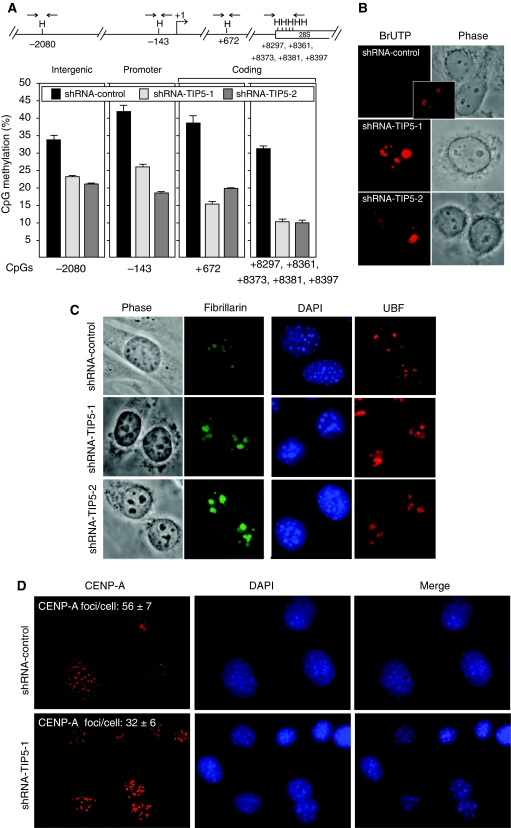
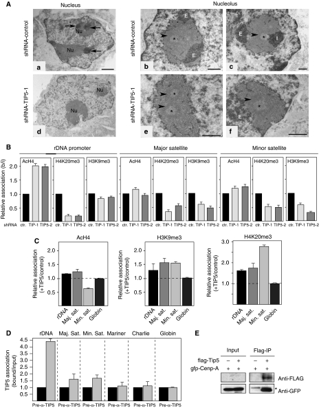
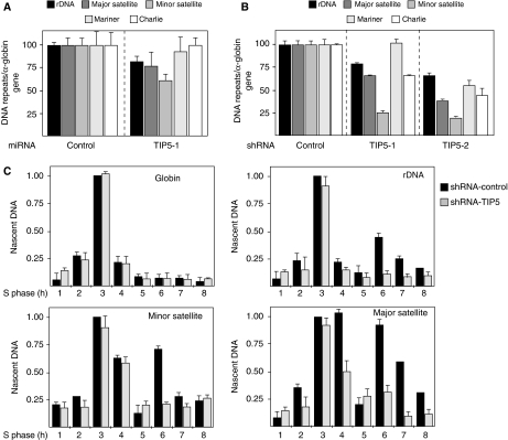
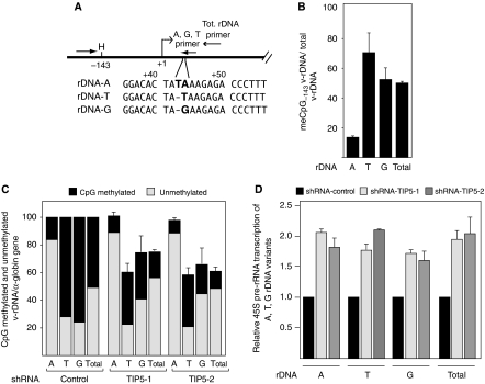
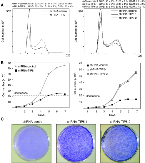
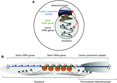
Maintenance of specific heterochromatic domains is crucial for genome stability. In eukaryotic cells, a fraction of the tandem-repeated ribosomal RNA (rRNA) genes is organized in the heterochromatic structures. The principal determinant of rDNA silencing is the nucleolar remodelling complex, NoRC, that consists of TIP5 (TTF-1-interacting protein-5) and the ATPase SNF2h. Here we showed that TIP5 not only mediates the establishment of rDNA silencing but also the formation of perinucleolar heterochromatin that contains centric and pericentric repeats. Our data indicated that the TIP5-mediated heterochromatin is indispensable for stability of silent rRNA genes and of major and minor satellite repeats. Moreover, depletion of TIP5 impairs rDNA silencing, upregulates rDNA transcription levels and induces cell transformation. These findings point to a role of TIP5 in protecting genome stability and suggest that it can play a role in the cellular transformation process.


Conflict of interest statement

The authors declare that they have no conflict of interest.

Figures

Figure 1
Figure 1
Depletion of TIP5 impairs perinucleolar heterochromatin formation. (A) Depletion of TIP5 decreases CpG methylation at the entire rDNA repeat. A schema representing a single mouse rDNA repeat and the analysed HpaII (H) sites. The arrows represent the primers used to amplify the HpaII-digested DNA. The data represent the amounts of HpaII-resistant rDNA normalized to the total rDNA calculated by amplification with primers encompassing DNA sequences lacking HpaII sites and undigested DNA. The error bars indicate the s.d. of three independent experiments. (B) Depletion of TIP5 enhances rDNA transcription. rRNA transcripts were detected by in situ BrUTP incorporation after same exposure time. The inset shows a longer exposure of one control cell. (C) Depletion of TIP5 alters the number and size of nucleoli. Indirect immunofluorescence analysis of the nucleolar protein fibrillarin (left panel) or UBF (right panel) in shRNA-TIP5 and control cells. (D) Indirect immunofluorescence analysis of shRNA-control and shRNA-TIP5-1 cells using anti-CENP-A antibodies. The values represent the average number of CENP-A-stained foci of 50 cells scored at random.
Figure 2
Figure 2
TIP5 mediates heterochromatin formation at major and minor satellites. (A) The distribution of the heterochromatin (CC) associated with the nucleoli of shRNA-control and shRNA-TIP5 cells. A general view of the nuclei (a, d) and the nucleoli (b, c, e, f). The contrast procedure reveals in dark the structures containing the nucleic acids, DNA or RNAs. (a) In the nucleus of an shRNA-control cell, the CC (arrows) are visible within the nucleolus (Nu) or at the nucleolar periphery. (d) In the nucleus of an shRNA-TIP5 cell, no CC are visible in or close to the nucleoli (Nu). (b, c) In shRNA-control nucleoli the CC are detected close (I, intra-CC) to the FC (*) or at the nucleolar periphery (E, extra-CC). (e, f) In shRNA-TIP5 nucleoli few CC are present (E). The arrowheads indicate the DFC (dense FC). Bar: (a, d)=1 μm; (b, c, e, f)=0.5 μm. (B) Depletion of TIP5 decreases repressive histone modification levels at the rDNA, major and minor satellite repeats. Quantitative ChIP analysis of cross-linked chromatin was precipitated with the indicated antibodies. The data are presented as the amounts of bound normalized to input and shRNA-control cell levels. The error bars indicate the s.d. of three independent experiments. (C) Overexpression of TIP5 modifies the heterochromatin of rDNA, major and minor satellites. The data are presented as a modified histone fold-change of NIH3T3 cells transiently transfected with TIP5-expression plasmids versus that in cells transfected with control plasmids. The error bars indicate the s.d. of two independent experiments. (D) ChIP showing association of TIP5 with a minor fraction of satellite repeats in NIH3T3 cells. The data are presented as the amounts of bound normalized to input and pre-immunoserum levels. The error bars indicate the s.d. of four independent experiments. (E) CENP-A interacts with TIP5 in vivo. HEK293T cells were co-transfected with GFP-tagged CENP-A plasmids in the presence and absence of pcDNA-FLAG-TIP5 and precipitated with anti-FLAG antibodies. Co-precipitated CENP-A was visualized on immunoblots using antibodies against GFP and FLAG. The signal indicated by the asterisk represents IgG. 10% of the lysate used for IP is shown (input). The low levels of FLAG-TIP5 in the input were below the detection limit.
Figure 3
Figure 3
TIP5 protects genome stability. (A) Depletion of TIP5 induces loss of rDNA, major and minor satellite repeats. qPCR of genomic DNA from NIH3T3 cells infected for 10 days with a retrovirus expressing miRNA-TIP5 and (B) from shRNA-TIP5 cells. The values were normalized to the amounts of α-globin genes and to control cells. The error indicate the s.d. of three independent experiments. (C) Depletion of TIP5 alters the replication timing profiles of rDNA, major and minor satellite repeats. Synchronized cells were pulse-labelled with BrdU in 1-h intervals and nascent DNA was immunoprecipitated using anti-BrU antibodies. To calibrate for DNA recovery during IP, BrdU-labelled E. coli DNA was added to the reactions. Nascent DNA was measured by qPCR. The values represent the amounts of immunoprecipitated DNA normalized to the amounts of BrdU-labelled β-lactamase gene. The error bars indicate the s.d. of two independent experiments.
Figure 4
Figure 4
Depletion of TIP5 induces loss of CpG methylated, silent rDNA repeats. (A) A schema representing rDNA polymorphisms at +43/+44 (A, G and T sequences). The arrows represent the primers used to specifically amplify v-rDNA. (B) The CpG methylation profile of the v-rDNA promoter region in NIH3T3 cells. Polymorphism-specific qPCR. The data represent the amounts of HpaII-resistant v-rDNA normalized to the corresponding total v-rDNA calculated by amplifications using primers encompassing v-DNA sequences lacking HpaII sites and undigested DNA. The error bars indicate the s.d. of three independent experiments. (C) TIP5 mediates the stability of silent rRNA genes. Polymorphism-specific qPCR of v-rDNA from shRNA-TIP5 and control cells. The data were normalized to the amounts of α-globin genes and to control cells. Silent, methylated rDNA represents the HpaII-resistent fraction relative to v-rDNA amounts. The HpaII-digested fraction corresponds to active genes, lacking CpG methylation. The error bars indicate the s.d. of three independent experiments. (D) Depletion of TIP5 enhances the transcription of active rRNA genes. rRNA transcripts originating from v-rDNA variants were measured by qRT–PCR. The data are presented as the amounts of v-rRNA transcripts normalized to GAPDH mRNA levels and to control cells. The error bars indicate the s.d. of four independent experiments.
Figure 5
Figure 5
Depletion of TIP5 induces cellular transformation. (A) FACS analysis of miRNA and shRNA-TIP5 cells. Data were quantified from two independent experiments. (B) The growth curve of miRNA-TIP5 and shRNA-TIP5 and control cells. Cellular confluence was reached at about day 5. Similar results were obtained in three independent experiments performed in triplicates. The error bar values of shRNA cells are hidden by symbols. (C) The transforming activity of TIP5 depletion. Cells were plated at low density on a 10-cm-diameter plate. After 14 days (8 days after confluence) cells were stained with methylene blue.
Figure 6
Figure 6
TIP5 mediates the heterochromatin at the nucleolar/perinucleolar associated chromatin. A model showing the role of TIP5 in establishing heterochromatin at regions located adjacent to the nucleolus. The cellular (A) and linear (B) distribution of active/silent rRNA genes and centromeric heterochromatin within the nucleolus and at the perinuclear periphery. In a transient association model, TIP5 interacts transiently and/or weakly with nearby localized chromatin domains (centric-pericentric repeats) from its stable binding sites (silent rRNA genes). Alternatively, spread of heterochromatin from silent rRNA genes or formation of nucleolar/perinucleolar compartment enriched in chromatin repressor complexes can affect the perinucleolar heterochromatin. In this model, it is also proposed that TIP5 and silent rRNA copies have a role in mediating the transcriptional activity of active rRNA genes as suggested by the results shown in Figure 4D.

References

    1. Akhmanova A, Verkerk T, Langeveld A, Grosveld F, Galiart N (2000) Characterisation of transcriptionally active and inactive chromatin domains in neurons. J Cell Sci 24: 4463–4474 - PubMed
    1. Arnheim N, Southern EM (1977) Heterogeneity of the ribosomal genes in mice and men. Cell 11: 363–370 - PubMed
    1. Black BE, Bassett EA (2008) The histone variant CENP-A and centromere specification. Curr Opin Cell Biol 20: 91–100 - PubMed
    1. Boisvert FM, van Koningsbruggen S, Navascués J, Lamond AI (2007) The multifunctional nucleolus. Nat Rev Mol Cell Biol 8: 574–585 - PubMed
    1. Carvalho C, Pereira HM, Ferreira J, Pina C, Mendonca D, Rosa AC, Carmo-Fonseca M (2001) Chromosomal G-dark bands determine the spatial organization of centromeric heterochromatin in the nucleus. Mol Cell Biol 12: 3563–3572 - PMC - PubMed

Publication types