. 2010 Mar 17;29(6):1081-90.
doi: 10.1038/emboj.2010.9. Epub 2010 Feb 18.

Bacterial actin MreB assembles in complex with cell shape protein RodZ

Affiliations

Bacterial actin MreB assembles in complex with cell shape protein RodZ

Fusinita van den Ent et al. EMBO J. .

Abstract

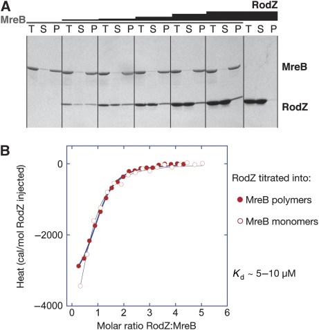
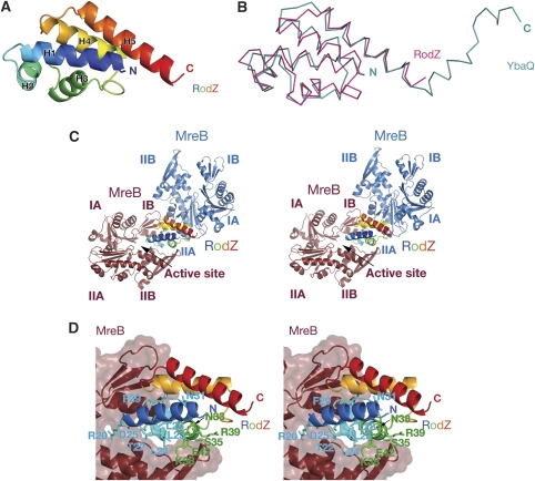
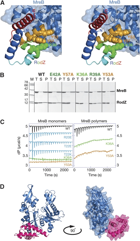
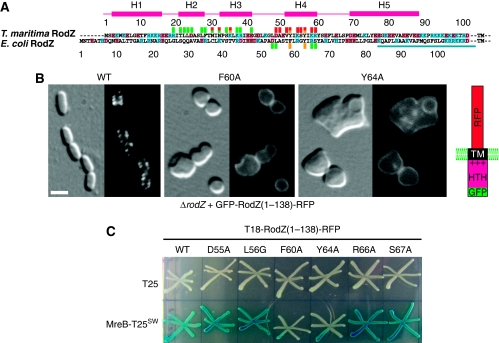
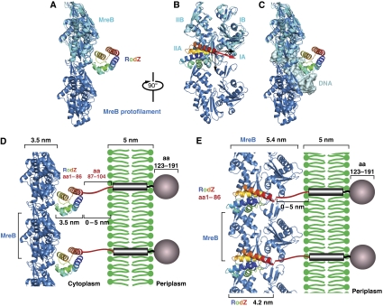
Bacterial actin homologue MreB is required for cell shape maintenance in most non-spherical bacteria, where it assembles into helical structures just underneath the cytoplasmic membrane. Proper assembly of the actin cytoskeleton requires RodZ, a conserved, bitopic membrane protein that colocalises to MreB and is essential for cell shape determination. Here, we present the first crystal structure of bacterial actin engaged with a natural partner and provide a clear functional significance of the interaction. We show that the cytoplasmic helix-turn-helix motif of Thermotoga maritima RodZ directly interacts with monomeric as well as filamentous MreB and present the crystal structure of the complex. In vitro and in vivo analyses of mutant T. maritima and Escherichia coli RodZ validate the structure and reveal the importance of the MreB-RodZ interaction in the ability of cells to propagate as rods. Furthermore, the results elucidate how the bacterial actin cytoskeleton might be anchored to the membrane to help constrain peptidoglycan synthesis in the periplasm.


Conflict of interest statement

The authors declare that they have no conflict of interest.

Figures

Figure 1
Figure 1
A direct interaction between purified cell shape proteins RodZ (1−104) and MreB from T. maritima. (A) MreB polymerisation assays. MreB (28 μM) was incubated at 37°C with nucleotide and MgCl2 in the absence (panel 1, lanes 1–3) and the presence of increasing amounts of RodZ (14 μM panel 2; 35 μM panel 3; 70 μM panel 4; 140 μM panel 5, 214 μM panel 6). In the absence of MreB, RodZ(1−104) remains in the supernatant (panel 7 with 280 μM RodZ). The reactions were centrifuged at 140 000 g and total (T), supernatant (S) and pellet (P) were analysed on a 10–20% SDS gel, stained with Coomassie. (B) Isothermal titration calorimetry shows RodZ(1−104) binds to monomeric and filamentous MreB with similar affinities. The cell contained 40 μM MreB and the syringe 1 mM RodZ(1−104), which was added over 18 injections of 2 uL. Before the experiments, proteins were dialysed to 20 mM Tris–HCl, pH 7.5, 1 mM EDTA, 1 mM sodium azide, 200 mM NaCl. The solid lines are the fit to the data and follow a simple binding model, showing a low micromolar affinity, exothermic binding enthalpy and 1:1 binding stoichiometry.
Figure 2
Figure 2
(A) Ribbon representation of the crystal structure of the cytoplasmic part of RodZ(2−88) from T. maritima at 2.9 Å resolution. The secondary structure elements are labelled according to their appearance in the primary sequence (N-terminus in blue, C-terminus in red). (B) Superposition of the structures of RodZ(2−88) (in magenta) and helix-turn-helix type putative transcriptional regulator YbaQ (pdb entry 2eby, in turquoise). The two proteins overlap with a root mean square deviation (rmsd) of 2.1 Å over 77 residues, Z-score 9.8 (using structural similarity search DALI (Holm and Sander, 1993). (C) Stereograph of a ribbon representation of the cocrystal structure of MreB and RodZ(2−88) shows two possible interfaces. A larger interface where RodZ binds in the active site cleft of MreB between subdomains IB and IIB (MreB in brown) and a smaller interface, engaging subdomain IIA of MreB (MreB in blue). RodZ(2−88) is coloured in rainbow colours with the N-terminus in blue and C-terminus in red. Although the crystal packing shows the regular packing of heterodimers, only one molecule of RodZ(2−88) is shown for clarity reasons. (D) Binding mode of RodZ(2−88) within the active site cleft of MreB in stereo, as observed in the crystals. Substitution of the residues of RodZ depicted as sticks do not affect MreB binding in solution according to isothermal titration calorimetry or polymerisation assays.
Figure 3
Figure 3
(A) Stereograph of RodZ's interaction with subdomain IIA of MreB (blue). Residues K36, Y53 and Y57, shown in spheres, are required for MreB interaction. (B) MreB polymerisation assays were used to test the interface mutants of RodZ for MreB binding. MreB (28 μM) was incubated with RodZ(1−104) (35 μM) in the presence of nucleotide and MgCl2 at 37°C and the pellet was separated from the supernatant by centrifugation at 140 000 g. Total (T), supernatant (S) and pellet (P) was analysed on a 10–20% gradient gel and stained with Coomassie. Mutations in RodZ(1−104) are shown above the panels in colours corresponding to the labelled residues in Figures 2D and 3A. Protein marker (M) depicts molecular weights of 78, 66, 42, 30, 17 and 12 kDa. (C) Isothermal titration calorimetry (ITC) confirmed the behaviour of the interface mutants. Wild-type (WT) or mutant RodZ (1−104) was titrated into a cell containing monomeric MreB (left panel) or filamentous MreB (right panel). Colour code as for Figures 2D and 3A. The cell contained 40 μM MreB and the syringe 1 mM RodZ, which was added over 18 injections of 2 μl. Before the experiments, proteins were dialysed to 20 mM Tris–HCl, pH 7.5, 1 mM EDTA, 1 mM NaN3, 200 mM NaCl. (D) Ribbon representation of the cocrystal structure of MreB (in blue) and RodZ(2−88) (in magenta) (left). A surface representation of the contacts between the two proteins is shown on the right, where the structure is rotated by 90°.
Figure 4
Figure 4
Colocalisation of purified proteins MreB and RodZ(1−104) from T. maritima. Alexa 555-labelled MreB polymers (red) were formed in the presence of crowding agent PEG 4000, ATPγS and MgCl2. Subsequently, Alexa 488-labelled RodZ(1−104) was added (green) (top panel). In the absence of PEG 4000, Alexa 555-labelled MreB (red) and Alexa 488-labelled RodZ(1−104) (green) colocalise as large structures from which filaments grow when mixed together in the presence of nucleotide and MgCl2 (middle panel). RodZ's mutation Y53A impairs MreB interaction and does not colocalise with the large MreB structures (lower panel). Left images taken with mCherry filter showing MreB alone, middle with YFP filter showing RodZ(1−104) alone, right panel merged image. Images were taken using a 100 × microscope oil immersion objective (Nikon) and a Photometrics CoolSNAP HQ2 camera. Typical exposures were 50–100 msec.
Figure 5
Figure 5
(A) Sequence alignment of RodZ cytoplasmic domain from T. maritima and E. coli. Numbers above the alignment refer to the T. maritima RodZ sequence, below the alignment to the E. coli sequence. TM refers to the transmembrane helix. Residues marked with a red square above the sequence contribute to the surface area buried by the interface between RodZ and MreB. Mutations in residues marked with a yellow square abolish MreB binding, whereas those marked with a green square do not interfere with MreB binding when mutated. The squares underneath the E. coli sequence indicate mutations that affect cell shape and MreB interaction in E. coli (yellow) or behave as the wild-type protein (green) (see Supplementary Table III). The bar (in turquoise) indicates the juxtamembrane region in E. coli RodZ. (B) The effect of interface mutations on RodZ1−138 localisation and its ability to impose rod shape on ΔrodZ cells in E. coli. Shown are ΔrodZ cells (FB60) expressing GFP-RodZ1−138-RFP (left panel), or a mutant version thereof (middle and right) from a construct integrated at the chromosomal attHK022 site. The middle and right panels show cells producing the F60A and Y64A version, respectively (corresponding to Y53 and Y57 in T. maritima RodZ, respectively). Note the inability of these cells to maintain rod-shape, as well as the even membrane distribution of fluorescence. Cells were grown in M9-mal with 250 μM IPTG to OD600=0.3–0.5, and were imaged live with DIC (left side of each panel) and GFP fluorescence (right side of each panel) optics. Bar equals 2 μ. The construct used, GFP-RodZ1−138-RFP, is shown in the cartoon on the right. (C) Effects of RodZ interface mutations on its in vivo interaction with MreB in E. coli. Strain BTH101 [cya-99] was cotransformed with plasmid pairs encoding the indicated T18- and T25-fusions, and individual colonies were patched on M9-agar containing 0.2% glucose, 40 mg/ml X-Gal and 250 μM IPTG. The plate was incubated at 30°C and imaged after 24 h. Plasmids used were the vector control pKNT25 [PlaclacZ′-t25] (upper row), pCH375 [PlacmreB′-t25-′mreB], encoding a sandwich fusion that carries the CyaA T25 domain inserted between helices 6 and 7 of MreB (Bendezú et al, 2009) (lower row), pCH395 [Plact18-rodZ(1–138)-rfp], encoding the CyaA T18 domain appended to the N-terminus of a version of RodZ in which its periplasmic domain has been substituted with mCherry (Bendezú et al, 2009) (column 1), and mutant derivatives of pCH395 (columns 2–7).
Figure 6
Figure 6
(A, B) Model of RodZ cytoplasmic domain in complex with an MreB protofilament. The crystal structure of the heterodimer of RodZ(1−88) (rainbow colours, N-terminus in blue, C-terminus in red) and MreB (cyan) was superimposed on the protofilament structure of MreB (blue) (van den Ent et al, 2001). The black, downwards-pointing arrow shows the MreB filament axis. The red arrow indicates the direction of the C-terminal helix of RodZ's cytoplasmic domain that is positioned at ∼20° of the normal to the filament axis. MreB subdomains IA, IB, IIA, IIB are labelled in cyan. (C) Model showing that RodZ's HTH domain is unable to bind MreB and the nucleoid simultaneously. The helix-turn-helix motif of RodZ is modelled onto a classical DNA-binding protein (phage 343 repressor, pdb entry 2OR1), revealing its hypothetical DNA-binding site. Superimposing this imaginary complex on to the heterodimer structure of MreB and RodZ(1−88) shows a steric clash between MreB and DNA. Colour code as in (B). Double-stranded DNA in grey (taken from pdb entry 2OR1). (D, E) Model showing how RodZ might anchor the bacterial actin cytoskeleton in the membrane. As the orientation of MreB relative to the membrane is not known, the MreB filaments could adopt any position between the two extremes depicted in (D, E), depending on the flexibility of the linker that connects the helix-turn-helix motif of RodZ to the transmembrane region. (D) The side-view shows RodZ(1−104) positioned between MreB and the membrane. In that case, the distance between MreB and the membrane could vary between ∼3.5 and 8.5 nm, depending on the orientation of the linker of RodZ. (E) Front-end view: the heterodimer between MreB and RodZ(1−104) is rotated by 90° relative to (D). In this model, the spacing between MreB and the membrane could be between 0 nm (if part of the linker folds back) and ∼4 nm (in case the linker is fully extended). As MreB would face the membrane with subdomain I, the lateral contacts between the protofilaments would be limited to subdomain II and/or the back of the molecule (E).

Similar articles

Cited by

References

    1. Adams PD, Grosse-Kunstleve RW, Hung LW, Ioerger TR, McCoy AJ, Moriarty NW, Read RJ, Sacchettini JC, Sauter NK, Terwilliger TC (2002) PHENIX: building new software for automated crystallographic structure determination. Acta Crystallogr D Biol Crystallogr 58: 1948–1954 - PubMed
    1. Aggarwal AK, Rodgers DW, Drottar M, Ptashne M, Harrison SC (1988) Recognition of a DNA operator by the repressor of phage 434: a view at high resolution. Science 242: 899–907 - PubMed
    1. Alyahya SA, Alexander R, Costa T, Henriques AO, Emonet T, Jacobs-Wagner C (2009) RodZ, a component of the bacterial core morphogenic apparatus. Proc Natl Acad Sci USA 106: 1239–1244 - PMC - PubMed
    1. Amos LA, van den Ent F, Löwe J (2004) Structural/functional homology between the bacterial and eukaryotic cytoskeletons. Curr Opin Cell Biol 16: 24–31 - PubMed
    1. Aravind L, Anantharaman V, Balaji S, Babu MM, Iyer LM (2005) The many faces of the helix-turn-helix domain: transcription regulation and beyond. FEMS Microbiol Rev 29: 231–262 - PubMed

Publication types

MeSH terms