Review
. 2010 Mar;10(3):181-93.
doi: 10.1038/nrc2809. Epub 2010 Feb 19.

Eicosanoids and cancer

Affiliations
Review

Eicosanoids and cancer

Dingzhi Wang et al. Nat Rev Cancer. 2010 Mar.

Abstract

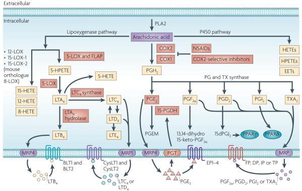
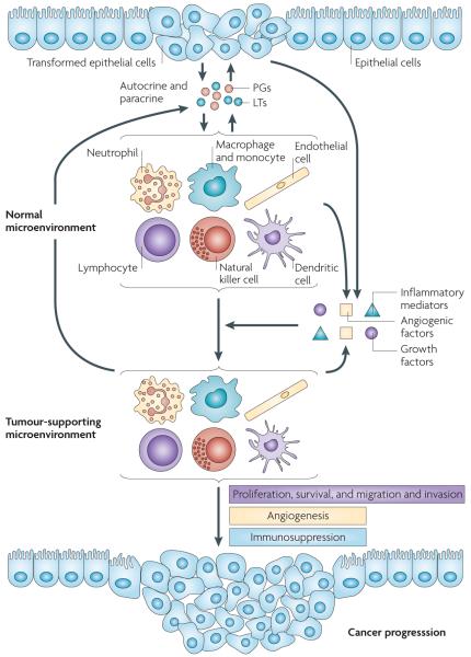
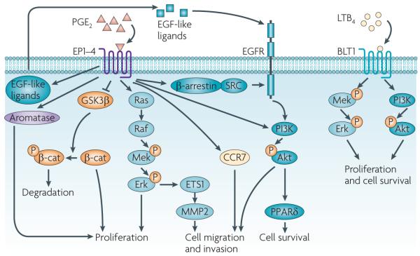
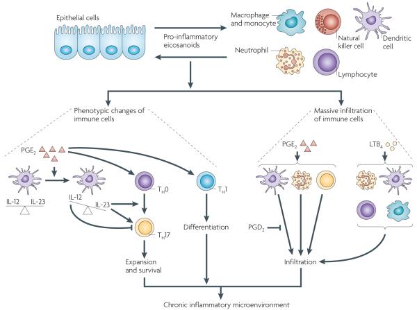
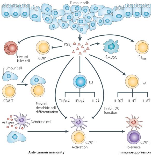
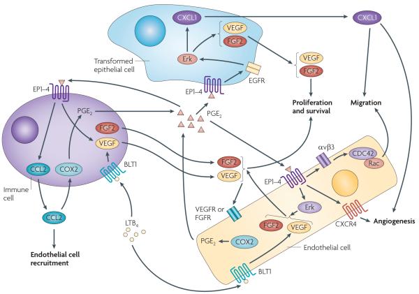
Eicosanoids, including prostaglandins and leukotrienes, are biologically active lipids that have been implicated in various pathological processes, such as inflammation and cancer. This Review highlights our understanding of the intricate roles of eicosanoids in epithelial-derived tumours and their microenvironment. The knowledge of how these lipids orchestrate the complex interactions between transformed epithelial cells and the surrounding stromal cells is crucial for understanding tumour evolution, progression and metastasis. Understanding the molecular mechanisms underlying the role of prostaglandins and other eicosanoids in cancer progression will help to develop more effective cancer chemopreventive and/or therapeutic agents.


Figures

Figure 1
Figure 1. An overview of eicosanoid synthesis pathways
Arachidonic acid is a polyunsaturated fatty acid that constitutes the phospholipid domain of most cell membranes and is liberated from the cellular membranes by cytoplasmic phospholipase A2 (PLA2). Free arachidonic acid can be metabolized to eicosanoids through three major pathways: the cyclooxygenase (COX), the lipoxygenase (LOX) and the cytochrome P450 monooxygenase pathways. In the COX pathway, the key step is the enzymatic conversion of arachidonic acid to the intermediate prostaglandin G2 (PGG2), which is then reduced to an intermediate PGH2 by the peroxidase activity of COX. PGH2 is sequentially metabolized to prostanoids, including prostaglandins (PGs) and thromboxanes (TXs) by specific prostaglandin and thromboxane synthases. LOXs convert arachidonic acid into biologically active metabolites such as leukotrienes and hydroxyeicosatetraenoic acids (HETEs); P450 metabolizes arachidonic acid into epoxyeicosatrienoic acids (EETs), HETEs and hydroperoxyeicosatetraenoic acids (HPETEs). In the 5-LOX pathway, arachidonic acid is converted to an intermediary 5-HPETE, which is further metabolized to form the unstable leukotriene A4 (LTA4). LTA4 is subsequently converted to 5-HETE, LTB4, LTC4, LTD4 and LTE4. Each of the prostaglandins and leukotrienes exerts its biological effects by binding to its cognate G protein-coupled receptor. PGI2 can transactivate the nuclear peroxisome proliferator-activated receptor-δ (PPARδ), and a PGD2 dehydration product, 15dPGJ2, is a natural ligand for PPARγ. The multidrug resistance-associated protein (MRP) gene family is comprised of efflux transporters for both prostaglandins and leukotrienes, and PGT is an influx transporter for prostaglandins. Hydroxyprostaglandin dehydrogenase 15-(NAD) (15-PGDH) mainly metabolizes intracellular PGE2 and PGF to a stable 13,14-dihydro-15-keto-PGE2 and 13,14-dihydro-15-keto-PGF. The red boxes indicate the signalling pathways that are discussed in this Review. CysLT, cysteinyl leukotriene; NSAID, non-steroidal anti-inflammatory drug.
Figure 2
Figure 2. Models of pro-inflammatory prostaglandins and leukotrienes in promoting cancer progression
Following the initiation of epithelial tumours, the reciprocal interactions between transformed epithelial and stromal cells have a key role in facilitating cancer progression. Pro-inflammatory prostaglandins and leukotrienes produced by tumour epithelial cells and their surrounding stromal cells are key mediators in this crosstalk and can accelerate tumour growth and metastasis through several methods. First, the pro-inflammatory prostaglandins (PGs) and leukotrienes (LTs) can directly induce epithelial tumour cell proliferation, survival, and migration and invasion in autocrine and paracrine manners. These pro-inflammatory lipids also stimulate epithelial cells and their surrounding stromal cells to produce growth factors, pro-inflammatory mediators and angiogenic factors, which switch the microenvironment from normal to tumour supporting. The tumour microenvironment, in turn, recruits immune cells and endothelial cells (tumour-infiltrating cells), which produce more pro-inflammatory mediators including eicosanoids, growth factors and angiogenic factors. These factors accelerate tumour growth and stimulate metastatsis through an autocrine loop by inducing angiogenesis and evading attack by the immune system.
Figure 3
Figure 3. PGE2 and LTB4 promote cancer progression through the induction of tumour epithelial cell proliferation, survival, and migration and invasion
Multiple cellular signalling pathways mediate the effects of prostaglandin E2 (PGE2) and leukotriene B4 (LTB4) on the regulation of epithelial tumour cell proliferation, survival, and migration and invasion. PGE2 stimulates cell proliferation through multiple cascades in both colorectal cancer (CRC) and non-small-cell lung cancer cells. PGE2 also induces cell proliferation through a glycogen synthase kinase-3β (GSK3β)–β-catenin (β-cat) pathway in CRC cells or by the upregulation of aromatase in breast cancer cells. PGE2 inhibition of GSK3β reduces β-catenin phosphorylation and thereby prevents its degradation, leading to accumulation, nuclear translocation and functional activity of β-catenin. PGE2 promotes CRC cell survival by activating a PI3K–Akt– peroxisome proliferator-activated receptor-δ (PPARδ) cascade in vitro and in vivo. In addition, PGE2 induces CRC cell migration and invasion through β-arrestin-1–SRC–epidermal growth factor receptor (EGFR)–PI3K–Akt signalling in vitro and in vivo. PGE2 transactivation of EGFR also depends on the extracellular release of an EGF-like ligand in CRC cell lines and a normal gastric epithelial cell line. PGE2 also induces cell migration and invasion through an Erk–ETS1–matrix metalloproteinase 2 (MMP2) cascade in pancreatic cancer cell lines or through the upregulation of C-C chemokine receptor 7 (CCR7) in breast cancer cell lines. LTB4 stimulates cell proliferation and promotes cell survival through a BLT1–Erk pathway in CRC cell lines or through both Mek–Erk and PI3K–Akt pathways in human pancreatic cancer cell lines. P, phosphorylation.
Figure 4
Figure 4. Prostaglandins and leukotrienes are key pro-inflammatory mediators in orchestrating crosstalk between tumour epithelial cells and immune cells
During the initiation of epithelial tumours or chronic inflammation, transformed or normal epithelial cells and tissue-resident immune cells locally secrete pro-inflammatory prostaglandins and leukotrienes such as prostaglandin E2 (PGE2) and leukotriene B4 (LTB4), which recruit large numbers of immune cells from the circulation into the mucosa and reprogramme the immune cells into pro-inflammatory leukocytes. For example, PGE2 induces expansion of inflammatory T helper 17 (TH17) cells by regulating interactions between T cells and dendritic cells and facilitates TH1 differentiation. PGE2 shifts the interleukin-12 (IL-12)/IL-23 balance in favour of IL-23 through the induction of IL-23 and inhibition of IL-12 expression in dendritic cells. IL-23 is essential for TH17 expansion and survival, whereas IL-12 suppresses TH17 development and function. In recruitment of immune cells, PGE2 induces infiltration of neutrophils and TH17 cells and enhances dendritic cell migration whereas PGD2 inhibits dendritic cell migration. LTB4 has a major role in attracting neutrophils, T cells, dendritic cells and macrophages from the circulation into inflammatory sites. Collectively, PGE2 and LTB4 induce the massive infiltration of immune cells and change their functionality, which in turn results in the establishment of a chronic inflammatory microenvironment.
Figure 5
Figure 5. PGE2 provides coordinated regulation of tumour immunosuppression
Pro-inflammatory prostaglandin E2 (PGE2) produced by tumour epithelial cells and/or their surrounding stromal cells induces immunosuppression through several ways, including: downregulating anti-tumour T helper 1 (TH1) cytokines and upregulating immunosuppressive TH2 cytokines; inhibiting CD8+ T cell proliferation and activity, suppressing the anti-tumour activity of natural killer cells and stimulating the expansion of regulatory T cells (TReg cells) and myeloid-derived suppressor cells (MDSCs); and inhibiting CD8+ T cell anti-tumour functions by impairing the ability of tumour cells to directly present tumour antigen, inhibiting dendritic cell differentiation and switching the function of dendritic cells from induction of immunity to T cell tolerance. The yellow CD8+ T cells have anti-tumour activity and the purple CD8+ T cell does not have anti-tumour activity. The purple dendritic cells have the ability to present tumour antigens from tumour cells with major histocompatibility complex (MHC) class I molecules to activate naive CD8+ T cells. The orange dendritic cell does not have the ability to activate CD8+ T cells (T cell tolerance). IFNγ, interferon-γ; IL, interleukin; TNFα, tumour necrosis factor-α.
Figure 6
Figure 6. A model of PGE2 and LTB4 coordinately regulating angiogenesis in the tumour microenvironment
Pro-inflammatory prostaglandin E2 (PGE2) and/or B4 LTB4 promote angiogenesis on at least two levels. First, PGE2 and/or LTB4 can directly act on epithelial, endothelial and/or immune cells to induce angiogenic factors such as vascular endothelial growth factor (VEGF), fibroblast growth factor 2 (FGF2) and the chemokines CXCL1 and CCL2. In transformed epithelial cells, PGE2 induces VEGF and CXCL1 secretion through an EP2 or EP4–epidermal growth factor (EGFR)–Erk cascade. In endothelial cells, PGE2 induces VEGF and FGF2 secretion through a MAPK pathway and LTB4 also stimulates VEGF expression. Moreover, PGE2 not only binds to endothelial cells to stimulate cell migration through an αVβ3 integrin–CDC42 and Rac pathway, but also mediates VEGF-and FGF2-induced CXCR4-dependent neovessel assembly in vivo. In immune cells, PGE2 promotes mast cells to release VEGF and CCL2, and LTB4 stimulates VEGF expression. Secretion of VEGF and FGF2 from tumour epithelial, endothelial and immune cells promotes endothelial cell proliferation and survival, and the chemokine CXCL1 released from tumour epithelial cells stimulates endothelial cell migration and tube formation in vitro and angiogenesis in vivo. CCL2 can attract endothelial cells to the tumour microenvironment. Interestingly, VEGF and FGF2 induce COX2 and subsequently PGE2 in endothelial cells, and CCL2 also induces COX2 and PGE2 in macrophages. Therefore, the effects of PGE2 on the regulation of VEGF, FGF2 and CCL2 are probably amplified through this positive feedback loop.

Similar articles

Cited by

References

    1. Vogelstein B, Kinzler KW. Cancer genes and the pathways they control. Nature Med. 2004;10:789–799. - PubMed
    1. Ting AH, McGarvey KM, Baylin SB. The cancer epigenome — components and functional correlates. Genes Dev. 2006;20:3215–3231. - PubMed
    1. Woutersen RA, Appel MJ, van Garderen-Hoetmer A, Wijnands MV. Dietary fat and carcinogenesis. Mutat. Res. 1999;443:111–127. - PubMed
    1. Harris RE. Cyclooxygenase-2 (cox-2) blockade in the chemoprevention of cancers of the colon, breast, prostate, and lung. Inflammopharmacology. 2009;17:55–67. - PubMed
    1. Pidgeon GP, et al. Lipoxygenase metabolism: roles in tumor progression and survival. Cancer Metastasis Rev. 2007;26:503–524. - PubMed

Publication types