. 2010 Feb 12;6(2):e1000790.
doi: 10.1371/journal.ppat.1000790.

Lethal antibody enhancement of dengue disease in mice is prevented by Fc modification

Affiliations

Lethal antibody enhancement of dengue disease in mice is prevented by Fc modification

Scott J Balsitis et al. PLoS Pathog. .

Abstract

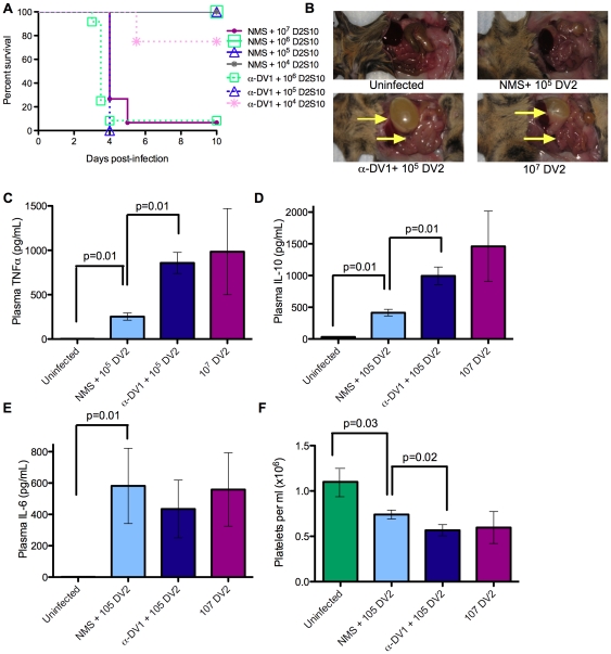
Immunity to one of the four dengue virus (DV) serotypes can increase disease severity in humans upon subsequent infection with another DV serotype. Serotype cross-reactive antibodies facilitate DV infection of myeloid cells in vitro by promoting virus entry via Fcgamma receptors (FcgammaR), a process known as antibody-dependent enhancement (ADE). However, despite decades of investigation, no in vivo model for antibody enhancement of dengue disease severity has been described. Analogous to human infants who receive anti-DV antibodies by transplacental transfer and develop severe dengue disease during primary infection, we show here that passive administration of anti-DV antibodies is sufficient to enhance DV infection and disease in mice using both mouse-adapted and clinical DV isolates. Antibody-enhanced lethal disease featured many of the hallmarks of severe dengue disease in humans, including thrombocytopenia, vascular leakage, elevated serum cytokine levels, and increased systemic viral burden in serum and tissue phagocytes. Passive transfer of a high dose of serotype-specific antibodies eliminated viremia, but lower doses of these antibodies or cross-reactive polyclonal or monoclonal antibodies all enhanced disease in vivo even when antibody levels were neutralizing in vitro. In contrast, a genetically engineered antibody variant (E60-N297Q) that cannot bind FcgammaR exhibited prophylactic and therapeutic efficacy against ADE-induced lethal challenge. These observations provide insight into the pathogenesis of antibody-enhanced dengue disease and identify a novel strategy for the design of therapeutic antibodies against dengue.


Conflict of interest statement

MSD has consulting agreements with MacroGenics, a company that has licensed the E60 monoclonal antibody from Washington University for possible commercial development. SJ is an employee of MacroGenics.

Figures

Figure 1
Figure 1. Lethal enhancement of dengue disease by anti-DV sera.
A. Mice were administered 100 µl naïve mouse serum (NMS) or anti-DV1 serum (α-DV1), challenged iv with the indicated dose of DV2 strain D2S10 24 hours later, and monitored for survival. Kaplan-Meier survival curves are shown; see Table S1 for p-values. The numbers of mice per group are as follows: NMS +107 D2S10, 15; NMS +106 D2S10, 8; NMS +105 D2S10, 11; NMS +104 D2S10, 4; α98J +106 D2S10, 12; α98J +105 D2S10, 4; α98J +104 D2S10, 4. B–F. Disease parameters were compared in mice receiving no virus, 107 pfu DV2, or 105 pfu DV2 after transfer of naïve or anti-DV1 serum. B. Vascular leak-associated fluid accumulation in visceral organs at day 3.5 post-infection. C–E. Serum levels of TNF-α (C), IL-10 (D), and IL-6 (E) in infected mice at day 3.5 post-infection were measured by ELISA (eBioscience). F. Platelet counts in mice at day 3.5 post-infection were determined using a hemocytometer. In (C–F), n = 4 in all groups except the uninfected group in panel F, where n = 12. Error bars represent standard deviations, and two-sided Wilcoxon rank sum tests were used to determine statistical significance.
Figure 2
Figure 2. Enhancement by heterologous anti-DV antibodies increases systemic viral burden of mouse-adapted and clinical isolates of DV.
A. Mice were administered 100 µl NMS or anti-DV1 serum ip and infected 24 hours later with 105 pfu DV2 D2S10 iv; lethal infection controls were infected iv with 107 pfu DV2 D2S10. Viral burden was measured in the indicated tissues at day 3.5 post-infection by qRT-PCR (serum) or plaque assay (all other tissues) as described in Materials and Methods. B. Mice were administered 100 µl NMS or anti-DV2 serum ip and infected the next day with 3×10⁶ pfu DV1 Western Pacific-74 iv. Virus burden was measured in the indicated tissues at day 3.5 post-infection by qRT-PCR (serum) or plaque assay (all other tissues) as described in the Materials and Methods C. Mice were administered 20 µg anti-DV MAb 4G2 or PBS ip and infected the next day with 106 pfu DV2 TSV01 iv. Virus burden was measured as in (A). Symbols indicate values in individual mice. Limits of detection are represented by dashed lines when present, or the horizontal axes. All pairwise comparisons were performed by two-sided Wilcoxon Rank Sum tests.
Figure 3
Figure 3. Detection and quantification of DV-infected cells with or without antibody-dependent enhancement.
Mice were administered naïve serum (NMS) or anti-DV1 serum and infected iv with 105 pfu DV2 the following day. Controls were mock-infected or infected with 107 pfu DV2 iv. A. Tissues were collected from all mice (n = 3–6 per group) at day 3.5, formalin-fixed, and processed into paraffin sections. Serial sections from each tissue were stained with anti-DV NS3 antibody E1D8 (NS3) or an isotype control mouse IgG2a (IgG2a data not shown), and multiple sections of each tissue type were thoroughly examined for staining. Positive staining for NS3 is brown while hematoxylin counterstain is blue. Strong cytoplasmic staining observed with E1D8, but not IgG2a control antibody, was considered DV-specific when observed in infected mice but not uninfected controls. NS3+ cells in lymph node, small intestine, and large intestine exhibited morphology and location consistent with tissue macrophages under all infection conditions (arrowheads). In liver, NS3+ cells were consistent with tissue macrophages and/or endothelial cells. Serial sections showed the F4/80 macrophage marker staining in the same locations where infected cells were present in lymph nodes, small intestine, large intestine, and bone marrow (data not shown). B. NS3+ cells per visual field were quantified. At least ten visual fields were counted for each sample except bone marrow, where four fields from four independent sections were counted due to the small area of mouse bone cross-sections. All pairwise comparisons were performed by two-sided Wilcoxon Rank Sum tests.
Figure 4
Figure 4. Antibody conditions for enhancement of DV infection.
A. Mice were administered 1.6–400 µl anti-DV1 serum ip, and pre-infection serum samples were collected the next day. Mice were then infected with 105 pfu DV2 iv and monitored for survival. Neutralizing activity of each pre-infection serum was determined in duplicate by PRNT50 assay on BHK21-15 cells. For each serum dose, PRNT50 results are displayed as the average of 3 to 4 mice, with the range in brackets. B. Serum transfers, bleeds, virus challenges, and PRNT assays were performed as in (A), but using anti-DV2 serum generated in AG129 mice. C. Viremia was measured in naïve serum controls (n = 4) and recipients of 400 µl anti-DV2 serum (n = 3) on day 4 post-infection by qRT-PCR. D. Mice were administered 1.25–400 µg of anti-DV monoclonal antibody 4G2, and pre-infection bleeds, challenges, and PRNT assays were performed as in (A).
Figure 5
Figure 5. Antibodies that lack the Fc region fail to mediate ADE and instead protect against lethal challenge.
A. Infection of K562 cells by DV2 in the presence of 4G2 mAb or 4G2 F(ab′)2 fragment was determined 48 hours post-infection by staining with Alexa488 anti-DV E protein MAb followed by flow cytometry. Average infection without antibody was 0.74%. B. Dosing scheme used to compare the in vivo effects of F(ab′)2 and intact mAb. Mice were administered intact 4G2 or IgG2a control mAb on day −1, or 20 µg doses of F(ab′)2 every 24 hours beginning 1 hour prior to infection, and were then challenged with 105 pfu of DV2 iv. C. Survival in mice from (B) receiving the indicated antibodies was scored on day 4 post-infection, and a two-sided Fisher's exact test was used. D. Viremia at day 4 post-infection in surviving mice from (C), measured by qRT-PCR. Error bars represent standard deviations, and pairwise comparisons were performed by two-sided Wilcoxon rank sum tests.
Figure 6
Figure 6. Antibodies with a mutated FcγR binding site cannot enhance DV infection in vitro or in vivo.
A. Infection of K562 cells by DV2 in the presence of E60-mIgG2a, E60-hIgG1, E60-N297Q. B. Mice were administered 20 µg of the indicated E60 mAbs, challenged 24 hours later with 106 pfu DV2 iv, and monitored for survival (n = 4 mice per group). p = 0.02 for E60-hIgG1 versus E60-N297Q. C. Mice were administered E60 mAbs and virus as in (B), and viral burden in peripheral blood cells was measured by plaque assay. n = 4 mice per group.
Figure 7
Figure 7. Antibodies with a mutated FcγR binding site have both prophylactic and therapeutic potential and reduce viral load and serum TNF-α in DV-infected mice.
A. Mice were simultaneously administered 25 µl anti-DV1 serum and 20 µg of the indicated E60 mAbs ip, challenged 24 hours later with 2×10⁵ pfu of DV2 iv, and monitored for survival (n = 5 mice per group). p = 0.009 for E60-hIgG1 versus E60-N297Q recipients. B. Mice were administered 25 µl anti-DV1 serum ip, challenged 24 hours later with 105 pfu of DV2 iv, treated by iv administration of E60-N297Q at the indicated doses and days post-infection, and monitored for survival. p-values compared to untreated controls (n = 9) were: p = 0.008 for 20 µg E60-N297Q on day 1 post-infection (n = 5); p = 0.005 for 20 µg E60-N297Q on day 2 post-infection (n = 10); and p = 0.02 for 50 µg E60-N297Q on day 2 post-infection (n = 5). Survival differences were compared using logrank tests. C. Mice were administered 25 µl of anti-DV1 serum ip and infected iv the next day with 105 pfu of DV2. Mice were injected iv with either PBS (untreated) or 20 µg E60-N297Q 24 hours later. On day 3.5 p.i, mice were euthanized and serum and tissues were collected. Viral burden in serum, small intestine, and bone marrow were measured by qRT-PCR for serum and plaque assay for solid tissues. D. Serum TNF-α was measured by ELISA. n = 4 mice per group in all analyses. For viral load and TNF-α values, error bars represent standard deviations, and pairwise comparisons were performed by two-sided Wilcoxon rank sum tests.

References

    1. Rothman AL. Dengue: defining protective versus pathologic immunity. J Clin Invest. 2004;113:946–951. - PMC - PubMed
    1. Halstead SB. Dengue. Lancet. 2007;370:1644–1652. - PubMed
    1. WHO . Geneva: World Health Organization; 1997. Dengue haemorrhagic fever: diagnosis, treatment, prevention, and control.
    1. Halstead SB. Neutralization and antibody-dependent enhancement of dengue viruses. Adv Virus Res. 2003;60:421–467. - PubMed
    1. Kliks SC, Nimmanitya S, Nisalak A, Burke DS. Evidence that maternal dengue antibodies are important in the development of dengue hemorrhagic fever in infants. Am J Trop Med Hyg. 1988;38:411–419. - PubMed

Publication types

Substances