

Molecular systematics of the marine Dothideomycetes
- PMID: 20169029
- PMCID: PMC2816972
- DOI: 10.3114/sim.2009.64.09
Molecular systematics of the marine Dothideomycetes
Abstract
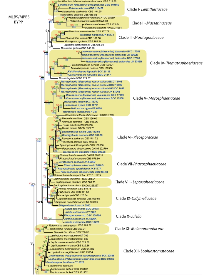
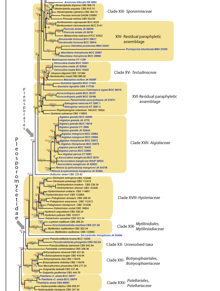
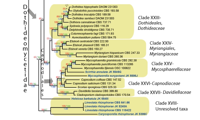
Phylogenetic analyses of four nuclear genes, namely the large and small subunits of the nuclear ribosomal RNA, transcription elongation factor 1-alpha and the second largest RNA polymerase II subunit, established that the ecological group of marine bitunicate ascomycetes has representatives in the orders Capnodiales, Hysteriales, Jahnulales, Mytilinidiales, Patellariales and Pleosporales. Most of the fungi sequenced were intertidal mangrove taxa and belong to members of 12 families in the Pleosporales: Aigialaceae, Didymellaceae,Leptosphaeriaceae, Lenthitheciaceae, Lophiostomataceae, Massarinaceae,Montagnulaceae, Morosphaeriaceae, Phaeosphaeriaceae, Pleosporaceae, Testudinaceae and Trematosphaeriaceae. Two new families are described: Aigialaceae and Morosphaeriaceae, and three new genera proposed: Halomassarina, Morosphaeria and Rimora. Few marine species are reported from the Dothideomycetidae (e.g. Mycosphaerellaceae, Capnodiales), a group poorly studied at the molecular level. New marine lineages include the Testudinaceae and Manglicolaguatemalensis in the Jahnulales. Significantly, most marine Dothideomycetes are intertidal tropical species with only a few from temperate regions on salt marsh plants (Spartina species and Juncus roemerianus), and rarely totally submerged (e.g. Halotthia posidoniae and Pontoporeia biturbinata on the seagrasses Posidonia oceanica and Cymodocea nodosum). Specific attention is given to the adaptation of the Dothideomycetes to the marine milieu, new lineages of marine fungi and their host specificity.
Keywords: Dothideomycetes; ecology; marine fungi; multi-locus; new genera; systematics.
Figures
Similar articles
-
Dothideomycetes and Leotiomycetes sterile mycelia isolated from the Italian seagrass Posidonia oceanica based on rDNA data.Springerplus. 2014 Sep 9;3:508. doi: 10.1186/2193-1801-3-508. eCollection 2014. Springerplus. 2014. PMID: 25279300 Free PMC article.
-
A molecular phylogenetic reappraisal of the Hysteriaceae, Mytilinidiaceae and Gloniaceae (Pleosporomycetidae, Dothideomycetes) with keys to world species.Stud Mycol. 2009;64:49-83S3. doi: 10.3114/sim.2009.64.03. Stud Mycol. 2009. PMID: 20169023 Free PMC article.
-
A multigene phylogeny of the Dothideomycetes using four nuclear loci.Mycologia. 2006 Nov-Dec;98(6):1041-52. doi: 10.3852/mycologia.98.6.1041. Mycologia. 2006. PMID: 17486979
-
Fungal Biodiversity in Salt Marsh Ecosystems.J Fungi (Basel). 2021 Aug 9;7(8):648. doi: 10.3390/jof7080648. J Fungi (Basel). 2021. PMID: 34436187 Free PMC article. Review.
-
Advances in taxonomy of genus phoma: polyphyletic nature and role of phenotypic traits and molecular systematics.Indian J Microbiol. 2014 Jun;54(2):123-8. doi: 10.1007/s12088-013-0442-8. Epub 2013 Dec 4. Indian J Microbiol. 2014. PMID: 25320410 Free PMC article. Review.
Cited by
-
Neodendryphiella, a novel genus of the Dictyosporiaceae (Pleosporales).MycoKeys. 2018 Jul 26;(37):19-38. doi: 10.3897/mycokeys.37.27275. eCollection 2018. MycoKeys. 2018. PMID: 30150880 Free PMC article.
-
Comparative mitochondrial genome analyses reveal conserved gene arrangement but massive expansion/contraction in two closely related Exserohilum pathogens.Comput Struct Biotechnol J. 2022 Mar 21;20:1456-1469. doi: 10.1016/j.csbj.2022.03.016. eCollection 2022. Comput Struct Biotechnol J. 2022. PMID: 35386100 Free PMC article.
-
Multigene phylogeny and taxonomy of Dendryphion hydei and Torula hydei spp. nov. from herbaceous litter in northern Thailand.PLoS One. 2020 Feb 5;15(2):e0228067. doi: 10.1371/journal.pone.0228067. eCollection 2020. PLoS One. 2020. PMID: 32023268 Free PMC article.
-
Dothideomycetes and Leotiomycetes sterile mycelia isolated from the Italian seagrass Posidonia oceanica based on rDNA data.Springerplus. 2014 Sep 9;3:508. doi: 10.1186/2193-1801-3-508. eCollection 2014. Springerplus. 2014. PMID: 25279300 Free PMC article.
-
The microbiome dynamics and interaction of endosymbiotic Symbiodiniaceae and fungi are associated with thermal bleaching susceptibility of coral holobionts.Appl Environ Microbiol. 2024 Apr 17;90(4):e0193923. doi: 10.1128/aem.01939-23. Epub 2024 Mar 6. Appl Environ Microbiol. 2024. PMID: 38445866 Free PMC article.
References
-
- Abdel-Wahab MA (2005). Diversity of marine fungi from Egyptian Red Sea mangroves. Botanica Marina 48: 348–355.
-
- Abdel-Wahab MA, Jones EBG (2000). Three new marine ascomycetes from driftwood in Australian sand dunes. Mycoscience 41: 379–388.
-
- Abdel-Wahab MA, Jones EBG (2003). Decaisnella formosa sp. nov. (Ascomycota, Massariaceae) from an Australian sandy beach. Canadian Journal of Botany 81: 598–600.
-
- Alias SA, Jones EBG (2000). Colonization of mangrove wood by marine fungi at Kuala Selangor mangrove stand, Malaysia. Fungal Diversity 5: 9–21.
-
- Alias SA, Jones EBG, Torres J (1999). Intertidal fungi from the Philippines, with a description of Acrocordiopsis sphaerica sp. nov. (Ascomycota). Fungal Diversity 2: 35–41.
LinkOut - more resources
Full Text Sources
Molecular Biology Databases