. 2010 Feb 16;5(2):e9228.
doi: 10.1371/journal.pone.0009228.

Vacuolar ATPase regulates surfactant secretion in rat alveolar type II cells by modulating lamellar body calcium

Affiliations

Vacuolar ATPase regulates surfactant secretion in rat alveolar type II cells by modulating lamellar body calcium

Narendranath Reddy Chintagari et al. PLoS One. .

Abstract

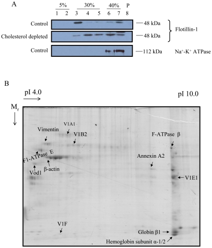
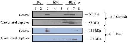
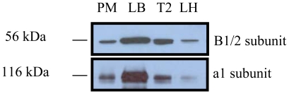
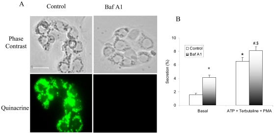
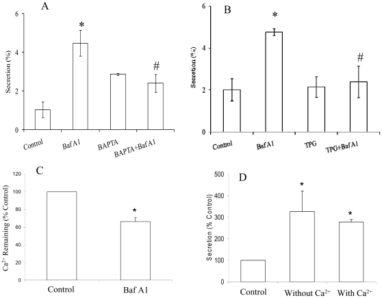
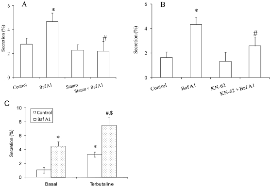
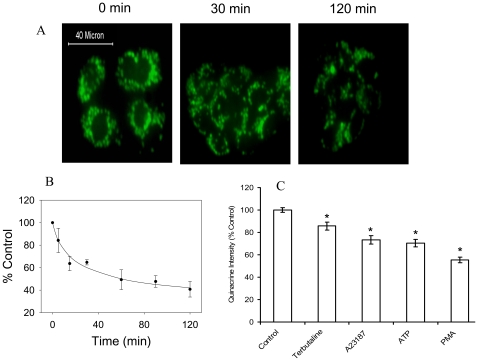
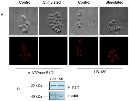
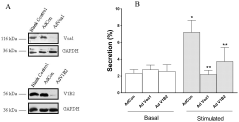
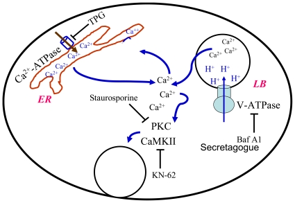
Lung surfactant reduces surface tension and maintains the stability of alveoli. How surfactant is released from alveolar epithelial type II cells is not fully understood. Vacuolar ATPase (V-ATPase) is the enzyme responsible for pumping H(+) into lamellar bodies and is required for the processing of surfactant proteins and the packaging of surfactant lipids. However, its role in lung surfactant secretion is unknown. Proteomic analysis revealed that vacuolar ATPase (V-ATPase) dominated the alveolar type II cell lipid raft proteome. Western blotting confirmed the association of V-ATPase a1 and B1/2 subunits with lipid rafts and their enrichment in lamellar bodies. The dissipation of lamellar body pH gradient by Bafilomycin A1 (Baf A1), an inhibitor of V-ATPase, increased surfactant secretion. Baf A1-stimulated secretion was blocked by the intracellular Ca(2+) chelator, BAPTA-AM, the protein kinase C (PKC) inhibitor, staurosporine, and the Ca(2+)/calmodulin-dependent protein kinase II (CaMKII), KN-62. Baf A1 induced Ca(2+) release from isolated lamellar bodies. Thapsigargin reduced the Baf A1-induced secretion, indicating cross-talk between lamellar body and endoplasmic reticulum Ca(2+) pools. Stimulation of type II cells with surfactant secretagogues dissipated the pH gradient across lamellar bodies and disassembled the V-ATPase complex, indicating the physiological relevance of the V-ATPase-mediated surfactant secretion. Finally, silencing of V-ATPase a1 and B2 subunits decreased stimulated surfactant secretion, indicating that these subunits were crucial for surfactant secretion. We conclude that V-ATPase regulates surfactant secretion via an increased Ca(2+) mobilization from lamellar bodies and endoplasmic reticulum, and the activation of PKC and CaMKII. Our finding revealed a previously unrealized role of V-ATPase in surfactant secretion.


Conflict of interest statement

Competing Interests: The authors have declared that no competing interests exist.

Figures

Figure 1
Figure 1. 2-D gel electrophoresis of alveolar type II cell lipid rafts.
(A) Confirmation of lipid rafts isolated from type II cells: Freshly isolated type II cells were lysed in the presence of 1% Triton X-100 (control) or 0.5% Triton X-100 and 0.5% saponin (cholesterol depleted) at 4°C for 45 min. Later, the lysate was subjected to a sucrose gradient centrifugation. Seven Fractions were collected from the top. The pellet was dissolved in lysis buffer (fraction P). The fractions were immunoblotted for flotillin-1, a raft marker protein and Na+-K+ ATPase, a non-raft marker protein. (B) 2-D gel electrophoresis: Lipid rafts proteins were subjected to 2-D gel electrophoresis. Later, the gels were stained with Coomassie blue. Shown is a representative of 3 runs.
Figure 2
Figure 2. Association of V-ATPase subunits with lipid rafts.
Type II cells were lysed in 1% Triton X-100 (control) or 0.5% Triton X-100+0.5% saponin (cholesterol depleted). Later, the lysates were subjected to raft isolation and various fractions were collected. Equal volumes of fractions were immunoblotted for V-ATPase B1/2 and a1 subunits.
Figure 3
Figure 3. Subcellular localization of V-ATPase subunits.
Total proteins isolated from plasma membrane (PM, 20 µg), lamellar bodies (LB, 5 µg), type II cells (T2, 20 µg), and lung tissue homogenates (LH, 20 µg) were immunoblotted for V-ATPase B1/2 and a1 subunits.
Figure 4
Figure 4. Effect of Bafilomycin A1 on the pH gradient across lamellar body membrane and surfactant secretion.
(A) Overnight cultured cells were treated with 20 nM Bafilomycin A1 (Baf A1) for 1 hr. The quinacrine was added to a final concentration of 10 µM. Fluorescence microscopy was undertaken to monitor quinacrine staining. Shown are representative images from 3 independent cell preparations. Scale bar: 10 µm. (B) Freshly isolated type II cells were labeled with [3H]choline overnight. The cells were treated with 20 nM Baf A1 for 1 hr, followed by incubation with a combination of 100 µM ATP; 10 µM terbutaline and 0.1 µM PMA for 2 additional hrs. Surfactant secretion was expressed as a percentage of total cellular [3H] PC secreted into medium. Data shown are means ± SE (n = 12). *P<0.05 v.s. Control (basal); #P<0.05 v.s. Baf A1 (basal); $P<0.05 v.s. Control (ATP+ Terbutaline +PMA) (ANOVA/Newman-Keuls Multiple Comparison Tests). Open bars, control; shaded bars, Baf A1.
Figure 5
Figure 5. Effects of intracellular Ca2+ on V-ATPase-mediated surfactant secretion.
(A, B) Overnight cultured type II cells were treated with 20 nM Bafilomycin A1 (Baf A1) in the presence or absence of the intracellular Ca2+-chelator, BAPTA-AM (50 µM) (Panel A) or the ER Ca2+-ATPase inhibitor, thapsigargin (TPG, 100 µM) (Panel B). Surfactant secretion was expressed as a percentage of total cellular [3H] PC secreted into medium. Data shown are means ± SE. *P<0.05 v.s. control; #P<0.05 v.s. Baf A1 (ANOVA/Newman-Keuls Multiple Comparison Tests, n = 3 independent cell preparations for Panel A and n = 4 independent cell preparations for Panel B). (C) Isolated lamellar bodies were loaded with 35Ca2+. After treated with 20 nM Baf A1 for 10 min, 45Ca retained in lamellar bodies was measured. *P<0.05 v.s. control (n = 4 lamellar body preparations). (D) Type II cells were incubated with 20 nM Baf A1 for 1 hr in Ca2+-free or 2 mM Ca2+ buffer and surfactant secretion was measured. *P<0.01 v.s. control (n = 3 independent cell preparations).
Figure 6
Figure 6. Effects of protein kinase inhibitors on V-ATPase-mediated surfactant secretion.
Overnight cultured type II cells were treated with 20 nM Bafilomycin A1 (Baf A1) in the presence or absence of staurosporine (Stauro, 10 nM) (Panel A), KN-62 (10 µM) (Panel B) or 20 µM terbutaline (Panel C). PC secretion was assayed. Data shown are means ± SE. *P<0.05 v.s. Control; #P<0.05 v.s Baf A1 (ANOVA/Newman-Keuls Multiple Comparison Tests, n = 3 independent cell preparations for Panel A and n = 4 independent cell preparations for Panel B). *P<0.05 v.s. Control (basal); #P<0.05 v.s. Baf A1 (basal); $P<0.05 v.s. Control (Terbutaline) (ANOVA/Newman-Keuls Multiple Comparison Tests, n = 3–4 independent cell preparations for Panel C).
Figure 7
Figure 7. Effect of lung surfactant secretagogues on the lamellar body pH in alveolar type II cells.
Overnight cultured type II cells were stimulated with a combination of 200 µM ATP and 0.2 µM PMA for various times indicated in the figure. At the end of incubation, the cells were immediately fixed and examined under a fluorescence microscope. (A) Shown are the representative images indicating the changes in quinacrine accumulation as a function of time. Scale bar: 40 µm. (B) The intensity of quinacrine staining was quantified and expressed as a percentage of control. Shown are means ± SE (n = 3 independent cell preparations). (C) Overnight cultured type II cells were stimulated with 20 µM terbutaline, 0.2 µM A23187, 1 mM ATP, or 0.1 µM PMA for 2 hours. The cells were incubated with quinacrine as above. Quinacrine staining intensity was quantified and expressed as arbitrary units. Shown are means ± SE (n = 3 independent cell preparations).
Figure 8
Figure 8. Differential localization of V-ATPase B1/2 subunits following stimulation of cells with lung surfactant secretagogues.
(A) Overnight cultured cells were stimulated with a combination of secretagogues 200 µM ATP and 0.2 µM PMA for 2 hrs. Immunostaining was performed to detect the localization of V-ATPase B1/2 subunit. The cells were also stained for the LB-180 protein to check for any changes in the lamellar body membrane following stimulation. Shown are the representative images. Scale bar: 40 µm. (B) Equal amounts of the total proteins (25 µg) isolated from control (Con) and stimulated (Sti) cells as in Panel A were immunobloted for V-ATPase B1/2 subunit and reprobed with β-actin. Shown are the representative immunoblots from 4 independent cell preparations.
Figure 9
Figure 9. Effect of V-ATPase subunit a1 and B2 knockdown on surfactant secretion by alveolar type II cells.
(A) Freshly isolated type II cells were transduced without (Blank Con) and with adenoviruses (MOI: 100) containing irrevelant siRNA sequences (AdCon), or siRNAs targeted to a1 (AdVoa1) and B2 (AdV1B2) subunits for 5 days. Cell lysates were immunoblotted for a1 and B2 proteins. The blots were reprobed for GAPDH for confirming equal loading of protein. (B) Type II cells were cultured on air-liquid cultures system. The cells were transduced with adenoviruses at a MOI of 100 on day 2 and cultured for 5 more days. The cells were labeled with [3H]-choline on day 6 overnight, then stimulated with 1 mM ATP for 2 hrs and assayed for surfactant secretion. The results were expressed as a percentage of unstimulated cells transduced with AdCon. Data shown are means ± SE (n = 4 independent cell preparations).
Figure 10
Figure 10. Schematic representation of proposed events for V-ATPase-mediated lung surfactant secretion.
Following V-ATPase inhibition by Baf A1 or lung surfactant secretagogues, Ca2+ is mobilized from lamellar bodies. The small localized changes in Ca2+ concentration leads to the further release of Ca2+ from ER store. The global increase in intracellular Ca2+ concentration results in the activation of PKC and CaMKII and the increase in surfactant secretion. LB: lamellar bodies; ER: endoplasmic reticulum.

Similar articles

Cited by

References

    1. Chintagari NR, Jin N, Wang P, Narasaraju TA, Chen J, et al. Effect of cholesterol depletion on exocytosis of alveolar type II cells. Am J Respir Cell Mol Biol. 2006;34:677–687. - PMC - PubMed
    1. Salaun C, James DJ, Chamberlain LH. Lipid rafts and the regulation of exocytosis. Traffic. 2004;5:255–264. - PMC - PubMed
    1. Pelkmans L, Helenius A. Endocytosis via caveolae. Traffic. 2002;3:311–320. - PubMed
    1. Allen JA, Halverson-Tamboli RA, Rasenick MM. Lipid raft microdomains and neurotransmitter signalling. Nat Rev Neurosci. 2007;8:128–140. - PubMed
    1. Schuck S, Simons K. Polarized sorting in epithelial cells: raft clustering and the biogenesis of the apical membrane. J Cell Sci. 2004;117:5955–5964. - PubMed

Publication types

MeSH terms