. 2010 Feb 16;8(2):e1000312.
doi: 10.1371/journal.pbio.1000312.

BIN1 localizes the L-type calcium channel to cardiac T-tubules

Affiliations

BIN1 localizes the L-type calcium channel to cardiac T-tubules

Ting-Ting Hong et al. PLoS Biol. .

Abstract

The BAR domain protein superfamily is involved in membrane invagination and endocytosis, but its role in organizing membrane proteins has not been explored. In particular, the membrane scaffolding protein BIN1 functions to initiate T-tubule genesis in skeletal muscle cells. Constitutive knockdown of BIN1 in mice is perinatal lethal, which is associated with an induced dilated hypertrophic cardiomyopathy. However, the functional role of BIN1 in cardiomyocytes is not known. An important function of cardiac T-tubules is to allow L-type calcium channels (Cav1.2) to be in close proximity to sarcoplasmic reticulum-based ryanodine receptors to initiate the intracellular calcium transient. Efficient excitation-contraction (EC) coupling and normal cardiac contractility depend upon Cav1.2 localization to T-tubules. We hypothesized that BIN1 not only exists at cardiac T-tubules, but it also localizes Cav1.2 to these membrane structures. We report that BIN1 localizes to cardiac T-tubules and clusters there with Cav1.2. Studies involve freshly acquired human and mouse adult cardiomyocytes using complementary immunocytochemistry, electron microscopy with dual immunogold labeling, and co-immunoprecipitation. Furthermore, we use surface biotinylation and live cell confocal and total internal fluorescence microscopy imaging in cardiomyocytes and cell lines to explore delivery of Cav1.2 to BIN1 structures. We find visually and quantitatively that dynamic microtubules are tethered to membrane scaffolded by BIN1, allowing targeted delivery of Cav1.2 from the microtubules to the associated membrane. Since Cav1.2 delivery to BIN1 occurs in reductionist non-myocyte cell lines, we find that other myocyte-specific structures are not essential and there is an intrinsic relationship between microtubule-based Cav1.2 delivery and its BIN1 scaffold. In differentiated mouse cardiomyocytes, knockdown of BIN1 reduces surface Cav1.2 and delays development of the calcium transient, indicating that Cav1.2 targeting to BIN1 is functionally important to cardiac calcium signaling. We have identified that membrane-associated BIN1 not only induces membrane curvature but can direct specific antegrade delivery of microtubule-transported membrane proteins. Furthermore, this paradigm provides a microtubule and BIN1-dependent mechanism of Cav1.2 delivery to T-tubules. This novel Cav1.2 trafficking pathway should serve as an important regulatory aspect of EC coupling, affecting cardiac contractility in mammalian hearts.


Conflict of interest statement

The authors have declared that no competing interests exist.

Figures
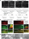
Figure 1
Figure 1. BIN1 colocalizes with Cav1.2 at T-tubules in cardiomyocytes.
(A) Confocal image (100×) of human (left) and mouse (right) adult cardiomyocytes. The cells were fixed and stained with mouse anti-BIN1 or rabbit anti-Cav1.2. Two-dimensional frames of Cav1.2 and BIN1 are shown in the top panel. Cardiomyocyte fluorescence intensity profiles along the cardiomyocyte longitudinal axis are presented in the middle panel. The bottom panel is the power spectrum over spatial distance averaged from five cardiomyocytes, which indicate that both BIN1 and Cav1.2 signals occurs at every 2 µm (fundamental peak occurs at ∼2 µm). Note the small peak at 1 µm is a harmonic of the fundamental peak at 2 µm (scale bar: 10 µm). (B) Confocal images (100×) of human (left) and mouse (right) cardiomyocytes stained with mouse anti-BIN1 (green) and rabbit anti-Cav1.2 (red) reveal colocalization between BIN1 and Cav1.2 along T-tubules (scale bar: 5 µm). Pearson colocalization coefficient and scatter plot between BIN1 and Cav1.2 are also shown in this panel. (C) Electron microscopy image of adult mouse cardiomyocytes fixed and immunogold labeled for BIN1 (small dots) and Cav1.2 (large dots) (scale bar: 200 nm) (left). As seen in the enlarged image, BIN1 and Cav1.2 occurs within 50 nm on T-tubule membranes. The negative control image without primary antibodies incubation is shown at the right panel.
Figure 2
Figure 2. BIN1 tethers dynamic microtubules.
(A) HeLa cells were transfected with α-Tubulin-GFP and BIN1-mCherry. The overlay pictures of BIN1 (red) and microtubules (black) are shown in the left panel. The right image is an enlarged subsection of the left image. Three microtubule travel paths (MT1, MT2, and MT3) are also highlighted in green in the subsection. (B) Graphs of each microtubule travel path. BIN1 edge (within 0.2 µm of BIN1 structure) is highlighted with a red dotted line in each graph.
Figure 3
Figure 3. Antegrade trafficking of Cav1.2 is microtubule dependent.
(A) Surface biotinylation of adult mouse cardiomyocytes indicates that nocodazole (30 µM) progressively reduces surface Cav1.2 expression in the presence of an endocytosis inhibitor dynasore (20 µM). Note that dynasore alone significantly increases surface expression of Cav1.2 by blocking dynamin-dependent endocytosis of Cav1.2 in cardiomyocytes. (B) Top panel: Confocal images (100×) of mouse cardiomyocytes stained with rabbit anti-Cav1.2 (red) and mouse anti-α-tubulin (green) reveal localization of Cav1.2 on microtubule network (scale bar: 5 µm). Bottom panel: Deconvolution of wide-field image of HL-1 cells stained with Cav1.2 (red) and α-tubulin (green). Merged image shows localization of Cav1.2 to the microtubule network. Enlarged pictures (right) indicate that Cav1.2 is distributed along microtubules (## p<0.01 when compared to control group, * p<0.05, ** p<0.01, when compared to vehicle group, Student's t test).
Figure 4
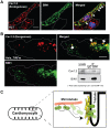
Figure 4. Cav1.2 is targeted to BIN1-induced membrane structures.
(A) Deconvolution of wide-field image (100×) of BIN1 transfected HL-1 cells indicates endogenous Cav1.2 (red) colocalizes with exogenous BIN1 (green) (scale bar: 5 µm). (B) TIRFm images of a HeLa cell transfected with Cav1.2-GFP (red) and BIN1-mCherry (green) reveal colocalization between BIN1 and Cav1.2 at the cell periphery (scale bar: 5 µm). This panel also includes co-immunoprecipitation between overexpressed BIN1-V5 (IP) and Cav1.2 (IB) in HeLa cells. (C) A schematic of dynamic microtubules delivering Cav1.2 to BIN1 at T-tubules.
Figure 5
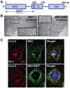
Figure 5. Cav1.2 is targeted to BIN1, not membrane invaginations.
(A) Domain map of wild-type BIN1 (BIN1). BIN1-BAR* (1-282 aa) contains the BAR domain and the sequence upstream of the coiled-coil region that is necessary for inducing membrane invagination. (B) Electron microscopy images indicate that BIN1 and BIN1-BAR* form similar membrane invaginations (dark linear tubules). (C) Deconvolved wide-field image of HL-1 cells transfected with BIN1 or BIN1-BAR*(1-282 aa). Co-staining between endogenous Cav1.2 (red) with transfected exogenous BIN1 or BIN1-BAR* (green) indicates that Cav1.2 localizes to BIN1 structures but not BIN1-BAR* structures (scale bar: 5 µm).
Figure 6
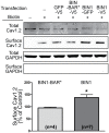
Figure 6. Full-length BIN1 causes Cav1.2 surface expression.
Surface biotinylation of Cav1.2 in HL-1 cells transfected with either BIN1 or BIN1-BAR* reveals that full-length BIN1 is required to cause surface expression of Cav1.2 (* p<0.05, Student's t test).
Figure 7
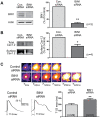
Figure 7. BIN1 knockdown delays calcium transient development in mouse cardiomyocytes.
(A) Western blot indicates an 80% knockdown of BIN1 protein by siRNA in differentiated mouse cardiomyocytes. (B) Surface biotinylation of Cav1.2 in these primary cardiomyocytes indicates a 45% reduction of surface Cav1.2 after BIN1 knockdown. (C) Live cell calcium imaging in differentiated cardiomyocytes indicates that BIN1 knockdown also delays calcium transient development in these cells. Average time to 50% maximal fluorescence intensity (T1/2 max) of calcium transient is presented in the left panel (* p<0.05, ** p<0.01, Student's t test).

Comment in

References

    1. Frost A, Unger V. M, De Camilli P. The BAR domain superfamily: membrane-molding macromolecules. Cell. 2009;137:191–196. - PMC - PubMed
    1. Butler M. H, David C, Ochoa G. C, Freyberg Z, Daniell L, et al. Amphiphysin II (SH3P9; BIN1), a member of the amphiphysin/Rvs family, is concentrated in the cortical cytomatrix of axon initial segments and nodes of ranvier in brain and around T tubules in skeletal muscle. J Cell Biol. 1997;137:1355–1367. - PMC - PubMed
    1. Frost A, Perera R, Roux A, Spasov K, Destaing O, et al. Structural basis of membrane invagination by F-BAR domains. Cell. 2008;132:807–817. - PMC - PubMed
    1. Lee E, Marcucci M, Daniell L, Pypaert M, Weisz O. A, et al. Amphiphysin 2 (Bin1) and T-tubule biogenesis in muscle. Science. 2002;297:1193–1196. - PubMed
    1. Ren G, Vajjhala P, Lee J. S, Winsor B, Munn A. L. The BAR domain proteins: molding membranes in fission, fusion, and phagy. Microbiol Mol Biol Rev. 2006;70:37–120. - PMC - PubMed

Publication types

MeSH terms