. 2010 Feb 12;6(2):e1000760.
doi: 10.1371/journal.ppat.1000760.

A novel pseudopodial component of the dendritic cell anti-fungal response: the fungipod

Affiliations

A novel pseudopodial component of the dendritic cell anti-fungal response: the fungipod

Aaron K Neumann et al. PLoS Pathog. .

Abstract

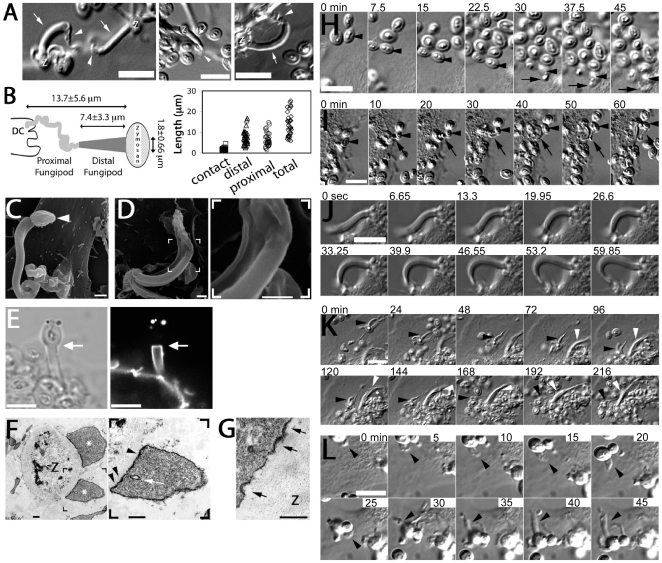
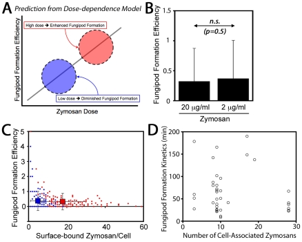
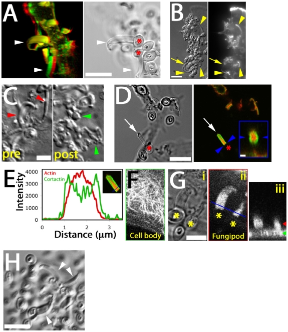
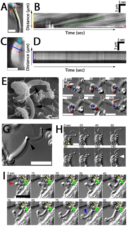
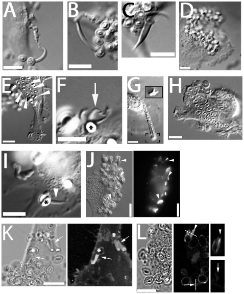
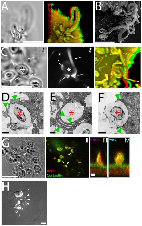
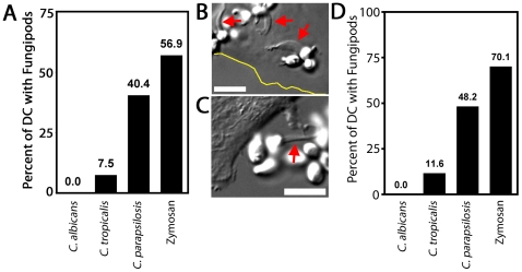
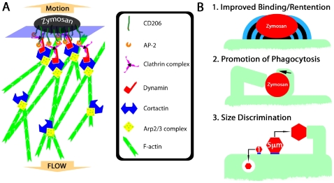
Fungal pathologies are seen in immunocompromised and healthy humans. C-type lectins expressed on immature dendritic cells (DC) recognize fungi. We report a novel dorsal pseudopodial protrusion, the "fungipod", formed by DC after contact with yeast cell walls. These structures have a convoluted cell-proximal end and a smooth distal end. They persist for hours, exhibit noticeable growth and total 13.7+/-5.6 microm long and 1.8+/-0.67 microm wide at the contact. Fungipods contain clathrin and an actin core surrounded by a sheath of cortactin. The actin cytoskeleton, but not microtubules, is required for fungipod integrity and growth. An apparent rearward flow (225+/-55 nm/second) exists from the zymosan contact site into the distal fungipod. The phagocytic receptor Dectin-1 is not required for fungipod formation, but CD206 (Mannose Receptor) is the generative receptor for these protrusions. The human pathogen Candida parapsilosis induces DC fungipod formation strongly, but the response is species specific since the related fungal pathogens Candida tropicalis and Candida albicans induce very few and no fungipods, respectively. Our findings show that fungipods are dynamic actin-driven cellular structures involved in fungal recognition by DC. They may promote yeast particle phagocytosis by DC and are a specific response to large (i.e., 5 microm) particulate ligands. Our work also highlights the importance of this novel protrusive structure to innate immune recognition of medically significant Candida yeasts in a species specific fashion.


Conflict of interest statement

The authors have declared that no competing interests exist.

Figures

Figure 1
Figure 1. Morphology and formation of fungipods.
(A) DIC images of representative fungipods on the dorsal surfaces of immature DC after 4 hours exposure to zymosan. “Z” denotes the location of the fungipod attached zymosan particle. Arrows and arrowheads designate distal and proximal fungipod regions, respectively. Bars = 10 µm. (B) Schematic of fungipod morphology and distribution of measured contact site widths as well as lengths of distal, proximal and total fungipod regions (N = 35). (C) SEM image of a typical fungipod with roughly cylindrical distal geometry (9500x). Arrowhead denotes the attached zymosan particle. Bar = 1 µm. (D) SEM image of a fungipod with ribbon-like distal geometry displaying longitudinal ridges. Right panel shows a higher magnification view of the flattened structure in the distal fungipod from the bracketed area of the left panel. Left panel, 7500x; Right panel, 25000x, Bar = 1 µm. (E) DiI-labeled membrane of a distal fungipod at the zymosan contact site (arrow) viewed as a medial confocal section of the distal fungipod. (left, DIC; right, confocal fluorescence). Bars = 5 µm. (F) A fungipod/zymosan contact site seen in thin section TEM imaging. The right panel displays the left panel bracketed region at higher magnification. Designations: “Z”, Zymosan; “*”, distal fungipod; arrow, example of vesicle inside distal fungipod; arrowheads, examples of pits/membrane densities at the contact site. Left panel, 2700x; Right panel, 6500x; Bars = 500 nm. (G) Thin section TEM of membrane invaginations at a zymosan contact site displaying studded juxtamembrane densities. 11000x, Bar = 500 nm. (H) DIC imaging of the initial attachment and nascence of a fungipod. Arrowheads and arrows designate a zymosan particle associated with fungipod generation and the nascent fungipod, respectively. Times indicate elapsed time of DC attachment for the indicated zymosan particle. Bar = 10 µm. (I) DIC imaging of the maturation of a fungipod. Arrowheads and arrows designate a relevant zymosan particle and the growing fungipod, respectively. Times indicate elapsed time since the advent of the fungipod. Bar = 10 µm. (J) DIC imaging of the growth of a mature fungipod. Bar = 10 µm. (K) DIC imaging of fungipods (arrowheads) associated with zymosan and DC for several hours. Bar = 10 µm. (L) DIC imaging of a fungipod (arrowhead) formed by a DC in response to live S. cerevisiae. Bar = 10 µm.
Figure 2
Figure 2. Zymosan dose-dependence of fungipods.
(A) Dose-dependence of fungipods predicts higher fungipod formation efficiencies at higher degrees of DC stimulation by zymosan particles. (B) Fungipod formation efficiency in DC-zymosan cultures is independent of zymosan density applied to DC. (n.s., not significant by Student's t-test) (C) Individual DC show no positive correlation between degree of zymosan stimulation and fungipod formation efficiency. For panels B & C, N = 165 cells for 20 µg/ml zymosan and N = 145 for 2 µg/ml zymosan. (D) There is no apparent correlation between degree of DC stimulation by zymosan and the kinetics of fungipod formation (N = 39 individual fungipod formation events).
Figure 3
Figure 3. Cytoskeletal structure of fungipods.
(A) Left panel: Red/green anaglyph of actin in DC fungipods (arrowheads) after 4 h exposure to zymosan. Non-fluorescent zymosan particles contacted the distal fungipod ends. Right panel: A single z-axis plane DIC image showing the position of zymosan particles (asterisk) with respect to fungipods (arrowhead). Bar = 10 µm. (B) Images of zymosan induced fungipods (arrowheads) in DIC (left panel) and actin-stained epifluorescence (right panel). The DIC image represents a single z-axis plane above the dorsal membrane, and actin is shown as a maximum intensity projection of all z-axis planes. Arrow indicates proximal fungipod. Bar = 10 µm. (C) DIC images of 4 hour zymosan-exposed DC fungipods before (“pre”) and after (“post”) treatment with cytochalasin D (15 minutes, 1 µm). Fungipods were well ordered before treatment (red arrowheads) and disordered after treatment (green arrowheads). Bar = 5 µm. (D) DIC (left panel) and confocal fluorescence imaging (right panel) of fungipods on DC after 4 hour zymosan exposure showing actin in red and cortactin in green at a fungipod-medial z-axis plane. Arrow and asterisk denotes fungipod and attached zymosan, respectively. Bar in left panel = 10 µm. Arrowheads in right panel designate the contact site and right panel inset is an orthogonal section through the contact site. Inset bar = 1 µm. (E) Linescan of actin (red) and cortactin (green) intensities (arbitrary units) in the distal fungipod. Location of line is shown in the inset image. (F) Confocal imaging of α-tubulin in the DC cell body showing microtubules. (G) (i) DIC image of fungipods at a medial z-axis plane with zymosan particles marked by asterisks. (ii) Diffuse α-tubulin staining in the distal fungipod. Blue line represents the region used for orthogonal section. (iii) Orthogonal section of previous panel showing fungipod diffuse tubulin in cross-section. Green and red arrowheads denote the z-axis positions of the cell body and fungipod images, respectively. Bar = 5 µm. (H) Fungipods (arrowheads) persist even after 1 hour exposure to nocodazol (10 µM). Bar = 10 µm.
Figure 4
Figure 4. Fungipod dynamics.
(A) Representative DIC image showing the zymosan particle (asterisk), contact site (arrowheads) and line used for kymograph analysis. Bar = 5 µm. (B) Representative kymograph showing DIC intensity along the line from the previous panel over time. Rearward flow appears as diagonal lines and speed of flow is measured from the slope of these lines. (C) DiI labeled distal fungipod overlaid with a line indicating the region used for kymography to observe changes in the fungipod edge position over time. Asterisk indicates position of attached zymosan. Bar = 5 µm. (D) Kymograph showing non-undulating edges of DiI stained distal fungipod. Arrowheads mark the edges of the fungipod. (E) SEM image of distal fungipods with arrows emphasizing the smooth lateral edges of these structures. Bar = 1 µm. (F) DIC time series showing rotation of a zymosan particle attached to a fungipod. The crosses and asterisks indicate the positions of the site of proximal fungipod attachment to the cell body and the zymosan particle, respectively. Bar = 10 µm. (G) DIC image of a fungipod showing a kinked appearance (arrowheads) developed during growth of the structure. Bar = 10 µm. (H) DIC time series showing supercoiling of a fungipod (arrowhead) attached to the zymosan particle indicated by an asterisk. Bar = 5 µm. (I) DIC time series showing a kinked fungipod (red arrowhead) associated with motion of the attached zymosan into the cell membrane as shown by the yellow reference line and green arrowhead. Blue arrowhead emphasizes relaxation of the kinked fungipod. Bar = 10 µm.
Figure 5
Figure 5. C-type lectin receptors and generation of fungipods.
(A) Laminarin (5 mg/ml) blocked DC produced normal fungipods. (B) Anti-dectin-1 polyclonal antibody (10 µg/ml) blocked DC produced normal fungipods. (C) Pharmacological inhibition of Dectin-1 signaling with Syk inhibitor II (1 µM) does not block formation of normal fungipods. (D) Blocking with soluble mannan (5 mg/ml) does inhibit the formation of fungipods although DC-associated zymosan particles are still observed. (E) Internalized zymosan particles (arrowheads) are still observed in the presence of blocking with soluble mannan. (F) Mannan-coated 5 µm beads (asterisk) induced protrusions (arrow) identical to fungipods. (G) Chitin particles induced protrusions identical to fungipods. Inset contains the bracketed area reproduced at 2× higher magnification than the main panel and focused at a lower plane to allow imaging of the chitin particle. (H) Blocking with anti-CD206 polyclonal antibody (50 µg/ml) abolished formation of zymosan induced fungipods but did not prevent binding to DC or internalization. (I) Anti-CD206 antibody coated 5 µm beads induced the formation of protrusions identical to fungipods. (J) DIC (left panel) and epifluorescence (right panel) imaging of intense CD206 staining at zymosan-DC contact sites (arrowheads). (K) DIC (left panel) and confocal fluorescence (right panel) imaging of clathrin light chain in zymosan (asterisks) induced fungipods. Right panel is a maximum intensity projection in the z-axis of a three dimensional confocal stack. Arrows indicate fungipods bearing clathrin LC. (L) DIC (left panel) and confocal fluorescence (middle and right panels) imaging of dynamin in DC/zymosan (asterisks) contacts. Lines denote locations of orthogonal sections through a membrane ruffle near a zymosan particle (arrowhead) and a mature fungipod (arrow). Orthogonal sections are shown in the right panels. Bar = 10 µm.
Figure 6
Figure 6. Functional significance of fungipods.
(A) DIC (left panel) and confocal 3D projection (right panel, red/green anaglyph) images of actin staining in DC with a zymosan particle attached in a cup-like structure formed by 14 fungipods. Zymosan position is shown in DIC. Bar = 10 µm. (B) SEM image (3700x) of zymosan particles with numerous attached fungipods. Bar = 10 µm. (C) DIC (left panel), confocal fluorescence (middle panel), and confocal 3D projection (right panel, red/green anaglyph) images of DiI stained DC with fungipods (arrows) enmeshing a zymosan particle (asterisk). Bar = 10 µm. (D) TEM thin section image of a zymosan particle (asterisk) being monolaterally engulfed (arrowheads) by a DC. Bar = 1 µm. (E) TEM thin section image of an internalized zymosan particle (asterisk) associated with overlapping phagocytic membranes (arrowheads). Bar = 1 µm. (F) TEM thin section image of a zymosan particle (asterisk) being bilaterally engulfed (arrowheads) by a DC. Bar = 1 µm. (G) DIC (i) and confocal fluorescence (ii) images of zymosan (asterisk) associated membrane wedges on DC (arrowheads) stained for actin (red) and cortactin (green). Orthogonal sections of a membrane wedge through horizontal (purple) and vertical (cyan) lines shown are depicted in panels (iii) and (iv), respectively. Bar = 10 µm for panels (i, ii). Bar = 1 µm for panels (iii, iv). (H) Representative DIC image of anti-CD206 coated 1 µm beads (arrowheads) bound to DC for 4 hours resulting in no fungipod-like protrusions. Bar = 10 µm.
Figure 7
Figure 7. Fungipods and Candida species.
(A) Percent of DC possessing surface-bound live yeast particles that made fungipods after 4 hour, 37°C DC co-culture with C. albicans (N = 82), C. tropicalis (N = 67), C. parapsilosis (N = 47) or zymosan (N = 65). (B) DIC image of C. parapsilosis on the dorsal surface of a DC (cell edge shown by yellow line) with associated fungipods (red arrows). (C) DIC image of C. tropicalis attached to a DC via a fungipod (red arrow). (D) Percent of DC possessing surface-bound fixed yeast particles that made fungipods after 4 hour, 37°C DC co-culture with C. albicans (N = 138), C. tropicalis (N = 129), C. parapsilosis (N = 114) or zymosan (N = 117). Bars = 10 µm.
Figure 8
Figure 8. Hypothetical mechanism of fungipod formation and functional roles of fungipods.
(A) Hypothetical protein complex involved in initiation and growth of fungipods. CD206 recruits clathrin complexes via AP-2 interactions with its tyrosine-based internalization motif. Clathrin binds dynamin, which can recruit cortactin leading to stabilization of Arp2/3 complexes and elongation of F-actin in dendritic networks. Cortactin also stabilizes actin branch points permitting greater longevity and stiffness of the F-actin network. Steady state polymerization of actin at the zymosan-proximal tip results in rearward flow of actin and a reaction force pushing the zymosan particle outward. (B) Summary of potential functional significances of fungipods for DC biology. (1) Fungipods may improve binding and retention of large particles such as zymosan that would be easily removed by shearing contact with other cells. (2) Fungipods may assist in phagocytosis (including monolateral engulfment/coiling phagocytosis) of yeast particles by holding this large particle in place during engulfment. (3) Fungipods may be part of the DC's specific size discriminatory response program directed against large (i.e., 5 µm) particles, but not smaller particles (i.e., 1 µm) that are more easily dealt with through conventional phagocytic means.

Similar articles

Cited by

References

    1. Banerjee SN, Emori TG, Culver DH, Gaynes RP, Jarvis WR, et al. Secular trends in nosocomial primary bloodstream infections in the United States, 1980–1989. National Nosocomial Infections Surveillance System. Am J Med. 1991;91:86S–89S. - PubMed
    1. Beck-Sague C, Jarvis WR. Secular trends in the epidemiology of nosocomial fungal infections in the United States, 1980–1990. National Nosocomial Infections Surveillance System. J Infect Dis. 1993;167:1247–1251. - PubMed
    1. Trofa D, Gacser A, Nosanchuk JD. Candida parapsilosis, an emerging fungal pathogen. Clin Microbiol Rev. 2008;21:606–625. - PMC - PubMed
    1. Lesage G, Bussey H. Cell wall assembly in Saccharomyces cerevisiae. Microbiol Mol Biol Rev. 2006;70:317–343. - PMC - PubMed
    1. Nguyen TH, Fleet GH, Rogers PL. Composition of the cell walls of several yeast species. Appl Microbiol Biotechnol. 1998;50:206–212. - PubMed

Publication types