

A broad distribution of the alternative oxidase in microsporidian parasites
- PMID: 20169184
- PMCID: PMC2820529
- DOI: 10.1371/journal.ppat.1000761
A broad distribution of the alternative oxidase in microsporidian parasites
Abstract
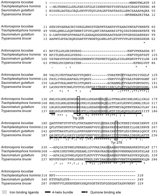
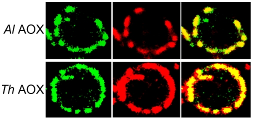
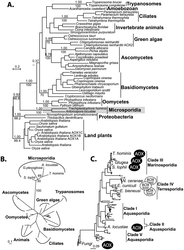
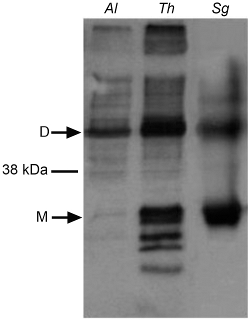
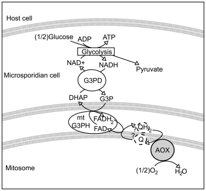
Microsporidia are a group of obligate intracellular parasitic eukaryotes that were considered to be amitochondriate until the recent discovery of highly reduced mitochondrial organelles called mitosomes. Analysis of the complete genome of Encephalitozoon cuniculi revealed a highly reduced set of proteins in the organelle, mostly related to the assembly of iron-sulphur clusters. Oxidative phosphorylation and the Krebs cycle proteins were absent, in keeping with the notion that the microsporidia and their mitosomes are anaerobic, as is the case for other mitosome bearing eukaryotes, such as Giardia. Here we provide evidence opening the possibility that mitosomes in a number of microsporidian lineages are not completely anaerobic. Specifically, we have identified and characterized a gene encoding the alternative oxidase (AOX), a typically mitochondrial terminal oxidase in eukaryotes, in the genomes of several distantly related microsporidian species, even though this gene is absent from the complete genome of E. cuniculi. In order to confirm that these genes encode functional proteins, AOX genes from both A. locustae and T. hominis were over-expressed in E. coli and AOX activity measured spectrophotometrically using ubiquinol-1 (UQ-1) as substrate. Both A. locustae and T. hominis AOX proteins reduced UQ-1 in a cyanide and antimycin-resistant manner that was sensitive to ascofuranone, a potent inhibitor of the trypanosomal AOX. The physiological role of AOX microsporidia may be to reoxidise reducing equivalents produced by glycolysis, in a manner comparable to that observed in trypanosomes.
Conflict of interest statement
The authors have declared that no competing interests exist.
Figures
Similar articles
-
Inhibition of mitosomal alternative oxidase causes lifecycle arrest of early-stage Trachipleistophora hominis meronts during intracellular infection of mammalian cells.PLoS Pathog. 2022 Dec 20;18(12):e1011024. doi: 10.1371/journal.ppat.1011024. eCollection 2022 Dec. PLoS Pathog. 2022. PMID: 36538568 Free PMC article.
-
Localization and functionality of microsporidian iron-sulphur cluster assembly proteins.Nature. 2008 Apr 3;452(7187):624-8. doi: 10.1038/nature06606. Epub 2008 Mar 2. Nature. 2008. PMID: 18311129
-
Microsporidian mitochondrial proteins: expression in Antonospora locustae spores and identification of genes coding for two further proteins.J Eukaryot Microbiol. 2005 May-Jun;52(3):271-6. doi: 10.1111/j.1550-7408.2005.05-00036.x. J Eukaryot Microbiol. 2005. PMID: 15927004
-
Back to basics: a revealing secondary reduction of the mitochondrial protein import pathway in diverse intracellular parasites.Biochim Biophys Acta. 2013 Feb;1833(2):295-303. doi: 10.1016/j.bbamcr.2012.02.006. Epub 2012 Feb 16. Biochim Biophys Acta. 2013. PMID: 22366436 Review.
-
Microsporidia: emerging pathogenic protists.Acta Trop. 2001 Feb 23;78(2):89-102. doi: 10.1016/s0001-706x(00)00178-9. Acta Trop. 2001. PMID: 11230819 Review.
Cited by
-
Re-identification of the ascofuranone-producing fungus Ascochyta viciae as Acremonium sclerotigenum.J Antibiot (Tokyo). 2017 Mar;70(3):304-307. doi: 10.1038/ja.2016.132. Epub 2016 Nov 2. J Antibiot (Tokyo). 2017. PMID: 27804952 No abstract available.
-
Molecular characterization and gene expression modulation of the alternative oxidase in a scuticociliate parasite by hypoxia and mitochondrial respiration inhibitors.Sci Rep. 2020 Jul 17;10(1):11880. doi: 10.1038/s41598-020-68791-9. Sci Rep. 2020. PMID: 32681023 Free PMC article.
-
Generation, Transfer, and Loss of Alternative Oxidase Paralogues in the Aspergillaceae Family.J Fungi (Basel). 2023 Dec 14;9(12):1195. doi: 10.3390/jof9121195. J Fungi (Basel). 2023. PMID: 38132795 Free PMC article.
-
Molecular Evolution of Alternative Oxidase Proteins: A Phylogenetic and Structure Modeling Approach.J Mol Evol. 2016 May;82(4-5):207-18. doi: 10.1007/s00239-016-9738-8. Epub 2016 Apr 18. J Mol Evol. 2016. PMID: 27090422
-
The Protean Acremonium. A. sclerotigenum/egyptiacum: Revision, Food Contaminant, and Human Disease.Microorganisms. 2018 Aug 16;6(3):88. doi: 10.3390/microorganisms6030088. Microorganisms. 2018. PMID: 30115839 Free PMC article.
References
-
- Keeling PJ, Doolittle WF. Alpha-tubulin from early-diverging eukaryotic lineages and the evolution of the tubulin family. Mol Biol Evol. 1996;13:1297–1305. - PubMed
-
- Edlind TD, Li J, Visvesvara GS, Vodkin MH, McLaughlin GL, et al. Phylogenetic analysis of beta-tubulin sequences from amitochondrial protozoa. Mol Phylogenet Evol. 1996;5:359–367. - PubMed
-
- Thomarat F, Vivares CP, Gouy M. Phylogenetic analysis of the complete genome sequence of Encephalitozoon cuniculi supports the fungal origin of microsporidia and reveals a high frequency of fast-evolving genes. J Mol Evol. 2004;59:780–791. - PubMed
Publication types
MeSH terms
Substances
Grants and funding
LinkOut - more resources
Full Text Sources
