Comparative Study
. 2010 Feb 12;6(2):e1000763.
doi: 10.1371/journal.ppat.1000763.

Pathogen entrapment by transglutaminase--a conserved early innate immune mechanism

Affiliations
Comparative Study

Pathogen entrapment by transglutaminase--a conserved early innate immune mechanism

Zhi Wang et al. PLoS Pathog. .

Abstract

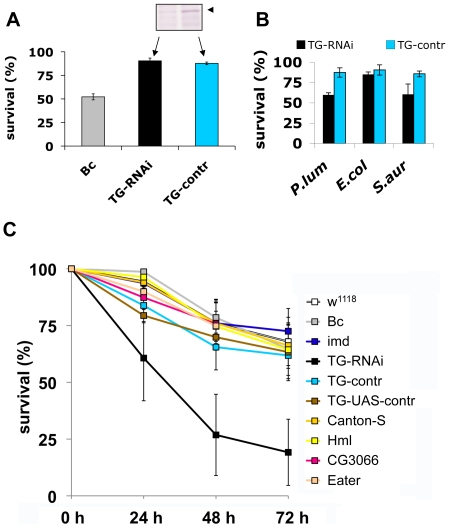
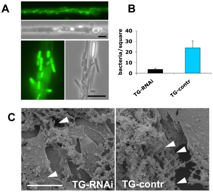
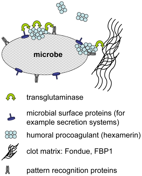
Clotting systems are required in almost all animals to prevent loss of body fluids after injury. Here, we show that despite the risks associated with its systemic activation, clotting is a hitherto little appreciated branch of the immune system. We compared clotting of human blood and insect hemolymph to study the best-conserved component of clotting systems, namely the Drosophila enzyme transglutaminase and its vertebrate homologue Factor XIIIa. Using labelled artificial substrates we observe that transglutaminase activity from both Drosophila hemolymph and human blood accumulates on microbial surfaces, leading to their sequestration into the clot. Using both a human and a natural insect pathogen we provide functional proof for an immune function for transglutaminase (TG). Drosophila larvae with reduced TG levels show increased mortality after septic injury. The same larvae are also more susceptible to a natural infection involving entomopathogenic nematodes and their symbiotic bacteria while neither phagocytosis, phenoloxidase or-as previously shown-the Toll or imd pathway contribute to immunity. These results firmly establish the hemolymph/blood clot as an important effector of early innate immunity, which helps to prevent septic infections. These findings will help to guide further strategies to reduce the damaging effects of clotting and enhance its beneficial contribution to immune reactions.


Conflict of interest statement

The authors have declared that no competing interests exist.

Figures

Figure 1
Figure 1. Drosophila Transglutaminase targets microbial surfaces.
Microbes analyzed include: yeast zymosan particles (A,B: Zym), Gram− E. coli (E.c.) and Gram+ S. aureus (S.a.; B) and the entomopathogenic nematode H. bacteriophora (C); A: phase contrast exposure (left), immunocytochemistry with an antibody against ε-(γ-glutamyl)lysine bridges created by TG (middle) and autofluorescence in the red channel (right). Note the punctate deposits (arrows) on the zymosan particles which are absent in preparations that lack hemolymph. B-cad was used in B and C, showing TG-mediated incorporation of B-cad into Gln-containing protein substrates. The inset at the upper left in B is a twofold enlargement to show the punctate labelling. All exposures were analyzed using immunofluorescence detecting B-cad and the corresponding phase contrast exposures. Hemolymph was omitted as a control leading to a reduction of the signal for all microbes. The fluorescence exposure of the reaction with zymosan lacking hemolymph (Fig. 1B, right part) was 10× overexposed to underline the significant difference in labelling efficiency between the presence or absence of hemolymph. After omission of B-cad signals were undetectable in most cases (not shown). Scale bars in A and B correspond to 5µm. C: Nematodes (Heterorhabditis bacteriophora) were incubated with B-cad and hemolymph leading to the formation of aggregates on the cuticle (upper part). The lower part shows autofluorescence after omission of hemolymph. The presence of GFP-expressing P. luminescens is indicated by arrows. The scale bar corresponds to 100µm.
Figure 2
Figure 2. Humoral procoagulants bind to microbial surfaces.
Lysates from E. coli (A) and S. aureus (B) were incubated in the presence of hemolymph (Hl), B-cad or the combination of both and analyzed using polyacrylamide gel electrophoresis. The additional band in the samples with Hl and B-cad (asterisk) represents hexamerin. Note that in the absence of B-cad hemolymph proteins form TG-crosslinked aggregates, thus preventing analysis with SDS-PAGE (see methods for further details). A similar pattern was obtained using P. luminescens (Figure S2). C: Proteins from an E. coli lysate treated like in Fig. 1 (right lane), were affinity-purified using streptavidin (Str-pure) and the identity of the purified proteins determined using mass spectrometry (E.c. shows a bacterial lysate, without hemolymph, the asterisks indicate breakdown products of hexamerin, see Table S1 for further details).
Figure 3
Figure 3. Human F XIII sequesters bacteria in the clot matrix.
A: Plasma obtained from healthy donors (FXIII +/+) or donors with FXIII-deficiency (FXIII −/−) was activated with thrombin in the presence of E. coli or S. aureus (for details see Methods). B-cad was used to visualize FXIII-mediated incorporation at bacterial surfaces by immunofluorescence microscopy. Samples were also visualized by phase contrast to show the contour of the bacteria. B: SEM exposures of clots formed with normal plasma or FXIII-deficient plasma (the insets correspond to an 8-fold higher magnification). Clots were formed in the absence of bacteria (ctrl) or the presence of E. coli or S. aureus (scale bars correspond to 5 µm in A and 10 µm in B).
Figure 4
Figure 4. F XIII crosslinks are detectable on bacterial surfaces.
E. coli (A–D) and S. aureus (A′–D′) bacteria were incubated with diluted and thrombin-activated normal (B–C′), F XIII-deficient plasma (D–D′) or left untreated (A–A′). Arrows in B and B′ point to plasma proteins crosslinked to the bacteria surface. Bacteria incubated with diluted and thrombin-activated normal (B–C′) or F XIII-deficient plasma (D–D′) were immunostained with a mouse anti-human gold-labeled ε-(γ-glutamyl) lysine-specific antibody. Arrowheads indicate crosslinking sites at the bacterial surface and arrows at crosslinking sites of crosslinked plasma proteins (scale bars correspond to 1 µm in B′ and 100 nm in D′). Please note that no colloidal gold staining was detected when bacteria were incubated with F XIII deficient plasma (D–D′).
Figure 5
Figure 5. Larvae with reduced TG levels show immune defects.
A: Lack of a wounding phenotype in TG knockdown lines. TG-knockdown larvae (Act5C-Gal4>UAS-TG-RNAi: labelled TG-RNAi); a control cross (Act5C-Gal4>w1118: labelled TG-control); and Bc larvae were injured and survival determined after 24 hours. The insert shows the reduction in TG protein levels (arrowhead) detected using TG-specific antibodies. B: TG knockdown larvae are more susceptible to some bacteria than control larvae. TG-RNAi larvae and control larvae were injected with P. luminescens, E. coli and S. aureus and survival scored after 24 h. C: TG-RNAi larvae are more susceptible to nematode infections. Larvae from the same strains like in (A) as well as the strain used for construction of knockdown lines (w1118); a homozygous imd mutant (imdY47); a wildtype strain (Canton-S) Hml mutants (Hml); mutants lacking CG3066 and eater mutants were infected with H. bacteriophora. Mortality rates were determined at the indicated times post-infection. All data points in Figs. A–C represent at least triplicates (+/− s.d.), all experiments were performed at 22°C, see Methods for further details.
Figure 6
Figure 6. Clots from larvae with reduced TG levels sequester fewer bacteria.
A: The clot from normal larvae captures P. luminescens. A clot bled from larvae expressing Fondue-FGP was drawn out from hemolymph in the presence of GFP-expressing P. luminescens as described . The clot is weakly labelled with Fondue-GFP . The bacteria, which are immobilised in the clot show a strong GFP signal (two sections are shown at different magnifications, the scale bars correspond to 10 µm). B: Hemolymph clots prepared as described from larvae with less TG (TG-RNAi) and control larvae were captured and the number of sequestered bacteria determined under the microscope (P<0.01, performed in triplicates). C: Clots from both types of larvae were also analyzed using scanning electron microscopy (note the more brittle appearance of the clot from TG-RNAi larvae, which is also observed after addition of MDC: see [14]). The scale bar corresponds to 10 µm, the arrowheads indicate bacteria which have been incorporated into the clot.
Figure 7
Figure 7. Hypothetical mechanisms for transglutaminase-mediated sequestration of microbes by the clot matrix.
Transglutaminase crosslinks humoral procoagulants such as hexamerin and Fondue leading to their incorporation into the clot. Additional possible TG-substrates on microbes include microbial surface proteins such as secretion systems and recognition proteins with specificity for microbial patterns.

Similar articles

Cited by

References

    1. Rittirsch D, Flierl MA, Ward PA. Harmful molecular mechanisms in sepsis. Nat Rev Immunol. 2008;8:776–787. - PMC - PubMed
    1. Sun H. The interaction between pathogens and the host coagulation system. Physiology (Bethesda) 2006;21:281–288. - PubMed
    1. Rowley AF, Ratcliffe NA. The granular cells of Galleria mellonella during clotting and phagocytic reactions in vitro. Tissue and Cell. 1976;8:437–446. - PubMed
    1. Bidla G, Lindgren M, Theopold U, Dushay MS. Hemolymph coagulation and phenoloxidase in Drosophila larvae. Dev Comp Immunol. 2005;29:669–679. - PubMed
    1. Scherfer C, Qazi MR, Takahashi K, Ueda R, Dushay MS, et al. The Toll immune-regulated Drosophila protein Fondue is involved in hemolymph clotting and puparium formation. Dev Biol. 2006;295:156–163. - PubMed

Publication types