. 2010 Feb 12;5(2):e9181.
doi: 10.1371/journal.pone.0009181.

Inheritance of DNA transferred from American trypanosomes to human hosts

Affiliations

Inheritance of DNA transferred from American trypanosomes to human hosts

Mariana M Hecht et al. PLoS One. .

Abstract

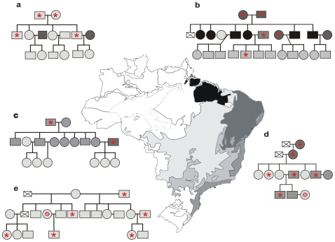
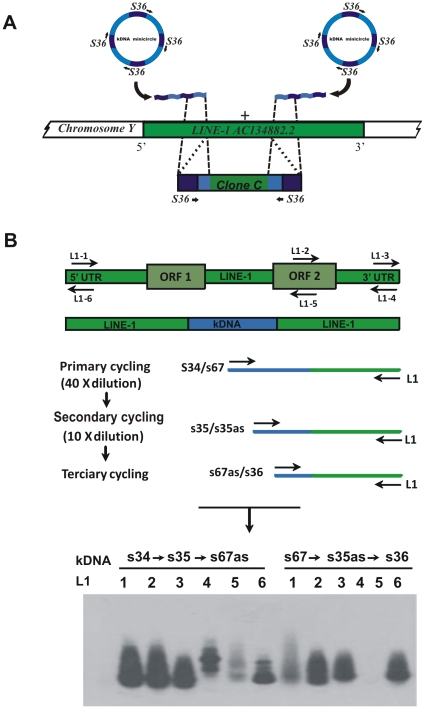
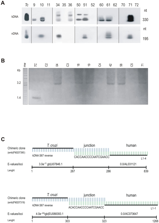
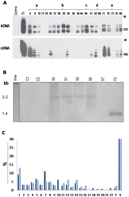
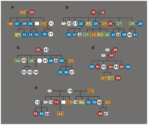
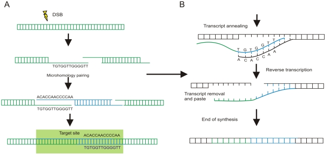
Interspecies DNA transfer is a major biological process leading to the accumulation of mutations inherited by sexual reproduction among eukaryotes. Lateral DNA transfer events and their inheritance has been challenging to document. In this study we modified a thermal asymmetric interlaced PCR by using additional targeted primers, along with Southern blots, fluorescence techniques, and bioinformatics, to identify lateral DNA transfer events from parasite to host. Instances of naturally occurring human infections by Trypanosoma cruzi are documented, where mitochondrial minicircles integrated mainly into retrotransposable LINE-1 of various chromosomes. The founders of five families show minicircle integrations that were transferred vertically to their progeny. Microhomology end-joining of 6 to 22 AC-rich nucleotide repeats in the minicircles and host DNA mediates foreign DNA integration. Heterogeneous minicircle sequences were distributed randomly among families, with diversity increasing due to subsequent rearrangement of inserted fragments. Mosaic recombination and hitchhiking on retrotransposition events to different loci were more prevalent in germ line as compared to somatic cells. Potential new genes, pseudogenes, and knockouts were identified. A pathway of minicircle integration and maintenance in the host genome is suggested. Thus, infection by T. cruzi has the unexpected consequence of increasing human genetic diversity, and Chagas disease may be a fortuitous share of negative selection. This demonstration of contemporary transfer of eukaryotic DNA to the human genome and its subsequent inheritance by descendants introduces a significant change in the scientific concept of evolutionary biology and medicine.


Conflict of interest statement

Competing Interests: The authors have declared that no competing interests exist.

Figures

Figure 1
Figure 1. Origin of family members in this study.
Map showing five main ecosystems in Brazil: light gray, Cerrado, savannah-like; intermediate gray, Inland Atlantic forest; gray, Coastal Atlantic forest; dark gray, Caatinga, dry shrubs; black, Amazon-Tocantins rain-forest. Family pedigrees a-to-e showing intermingling of human populations from the five main ecosystems. Based on their epidemiological histories, families a, b, and c live in the urban Federal District of Brazil, where individuals are not exposed to the insect transmitters of T. cruzi infection. By contrast, country families d and e live in houses infested with the cone-nosed kissing bugs, vectors of T. cruzi infection. The red asterisks indicate family members with chronic T. cruzi infection as detected by ELISA, hemagglutination, and immunofluorescence exams, and by the parasite DNA signature. The red disks indicate individuals showing the parasite DNA signature in absence of anti-T. cruzi antibodies.
Figure 2
Figure 2. The modified tpTAIL-PCR used to detect Trypanosoma cruzi minicircle integrations into the human genome.
(A) Truncated fragments of minicircles found in the LINE-1 copy at chromosome Y ; clone C showing conserved region annealed primer S36 (dark blue) at both ends followed by minicircle variable regions (light blue) and the LINE-1 sequence (green). (B) Targeted LINE-1 primers were used in combination with kDNA primer sets shown on top of the gel. The nested PCR reactions performed sequentially amplified target kDNA-host DNA sequences. The tertiary cycling products hybridizing with kDNA kCR probe on blots of 1% agarose gels were cloned and sequenced.
Figure 3
Figure 3. LkDT in somatic cells suggests mechanism of kDNA integration by homologous recombination.
(A) Representative PCR amplifications of T. cruzi kDNA and nDNA. kDNA and nDNA signatures indicate active infections and LkDTs, while only kDNA indicates LkDT. Amplification products obtained with kDNA (S35/S36) and nDNA (Tcz1/Tcz2) primer sets hybridized with specific internal probes S67 and Tcz3, respectively, on blots of 1% agarose gels. The numbers above each lane indicates the study case. Tc, T. cruzi. Patients 10, 34, 51, 61 and 71, showing nDNA and kDNA signatures, harbor active T. cruzi infections. Patients 9, 36, 50 and 60, with only the kDNA signature, may carry inherited integration events. (B) Southern hybridization of integrated kDNA revealed by a minicircle (kCR) probe. A 0.8% agarose gel was used to analize EcoRI-digested DNA from Chagas patient blood mononuclear cells. Top numbers indicate family founders who had the kDNA integrations in LINE-1 on chromosome X (Table S3). DNA from uninfected human donors (C2 to C5) and from T. cruzi were included as controls. (C) Schematic representation of microhomology-mediated end-joining kDNA minicircle integration into retrotransposon LINE-1. Patients 71 (top) and 24 (bottom) show AC-rich intermediates involved in recombination. Each patient showing nDNA and/or kDNA footprints yielded at least one chimeric sequence.
Figure 4
Figure 4. Tracking kDNA into germ line cells, and distribution of somatic and germline cells integrations into chromosomes.
(A) Representative PCR amplifications of T. cruzi kDNA and nDNA from male gametes, obtained with kDNA S35/S36 and nDNA Tcz1/Tcz2 primers, and hybridization to a blot of a 1% agarose gels with the kCR probe. Top numbers indicate study cases. nDNA and kDNA signatures indicate active infections and LkDTs, while kDNA indicate VkDT. (B) Southern blot hybridizations of EcoRI digestions of human DNA with a kDNA-specific probe on blots of 0.8% agarose gels. Top numbers indicate family c F1 progeny (patient 50) married to Chagas case patient (51) showing kDNA integrations in LINE-1 on chromosome X. The DNA from the parents and from two progeny (patients 55 and 56) formed 3.2-kb bands, whereas DNA from progeny 57 and from uninfected patients (C1 and C2) did not. Tc, T. cruzi. (C) Frequency of kDNA integrations in chromosomes of somatic (dark blue) and of germ-line (light blue) cells.
Figure 5
Figure 5. Co-localizing Trypanosoma cruzi kDNA sequence in metaphase plate chromosomes from human blood mononuclear cells.
kDNA integrations were co-localized in metaphase plates of nine patients from families a, b, and c with the 350-bp probe resulting from NsiI digest of wild-type kDNA, and with the LINE-1 clone 198 probe. (A) T. cruzi epimastigote fluorescein-labeled antibody (green) from Chagas patient serum (left) and control non-chagasic serum (right). The parasite silhouette is seen by digital image of contrast. B) T. cruzi DNA (Hoechst in blue), nDNA (Cy3 in red) and kDNA (FITC in green). The merged panel showed the parasite DNAs positioned with the digital image of contrast. C) Leishmania (viannia) braziliensis Hoechst stained DNA (blue); in absence of staining red (Cy3-LINE) and green (fluorescein-kDNA), respectively, with the 350-bp kDNA and with the LINE clone 180 probes. The merge was not positioned with the digital image of contrast. D) Metaphase chromosomes (blue), LINE-1 (red) and kDNA (green). The merge positioned with the digital image of contrast. Scale bars  = 5 µm.
Figure 6
Figure 6. Phylogenetic patchwork resulting from LkDT and VkDT.
Donors of diploid peripheral blood mononuclear cell DNA: Male, open square; female, open circle. Donors of haploid sperm cell DNA are indicated by yellow-edged square. Patients showing T. cruzi nDNA and kDNA are represented by red square or circle. Patients having integrated kDNA alone are identified by blue square or circle. Absence of a serial number means no biological sampling. A case deceased is indicated by crossed square. Volunteer samples were challenged by nDNA and kDNA amplifications withTcz1/Tcz2 and with S34/S67 primer sets, and blot hybridization with nDNA and kDNA probes. Each family member having nDNA and/or kDNA footprints yielded at least one chimeric sequence.
Figure 7
Figure 7. Model of Trypanosoma cruzi minicircle integration and replication in the human genome.
(A) Infection-induced host DNA (green) double strand-break (DSB) and integration of kDNA minicircle sequence (blue) mediated by microhomology end-joining. (B) Replication of the kDNA-LINE sequence by target-primed reverse-transcription using the autonomous recombination machinery of the LINE-1. The kDNA sequence repeat received a specific cut, and free-end pairing with a first strand transcript (green). A complementary second strand transcript is made, which can transpose to different chromosomes.

Similar articles

Cited by

References

    1. WHO Control of Chagas disease: second report of a WHO Expert Committee. 2002. pp. 1–109.
    1. Teixeira AR, Nitz N, Guimaro MC, Gomes C, Santos-Buch CA. Chagas disease. Postgrad Med J. 2006;82:788–798. - PMC - PubMed
    1. Teixeira AR, Nascimento R, Sturm NR. Evolution and Pathology in Chagas disease. Mem Inst Oswaldo Cruz. 2006;101:463–491. - PubMed
    1. Teixeira AR. Clinic Presentation of Chagas Disease. In: Teixeira A, Vinaud MC, Castro AM, editors. Emerging Chagas Disease. Bentham Science Publishers; 2009. pp. 104–109. : eISBN 978-1-60805-041-3.
    1. Teixeira AR, Figueiredo F, Rezende Filho J, Macedo V. Chagas disease: a clinical, parasitological and immunological study in rabbits. Am J Trop Med Hyg. 1983;32:258–272. - PubMed

Publication types

MeSH terms

Associated data