

Three-dimensional high-resolution reconstruction of the human gastro-oesophageal junction
- PMID: 20169612
- PMCID: PMC4106913
- DOI: 10.1002/ca.20941
Three-dimensional high-resolution reconstruction of the human gastro-oesophageal junction
Abstract
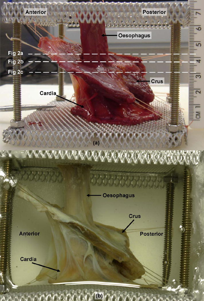
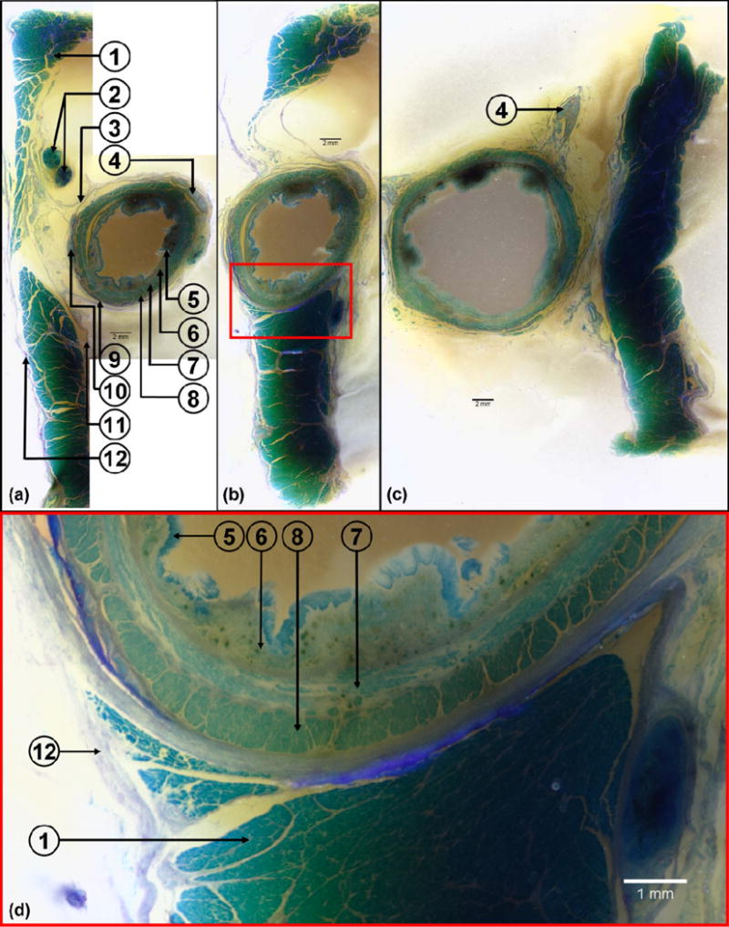
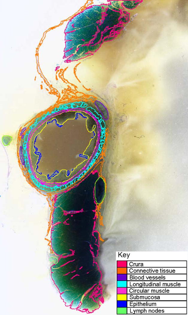
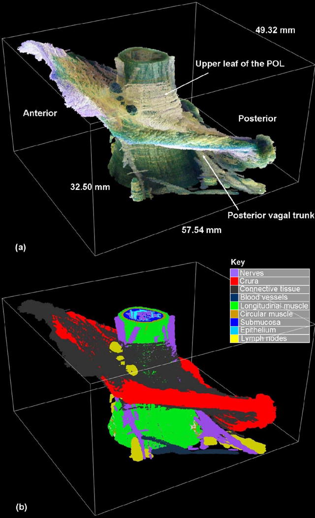
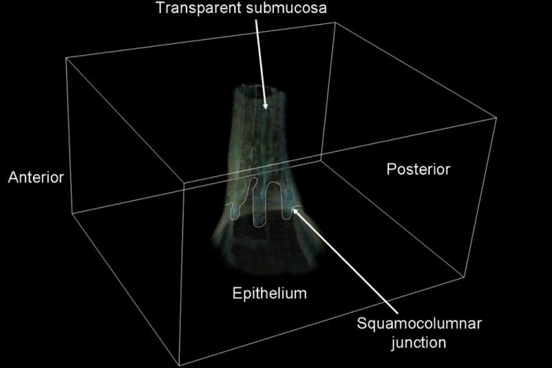
The aim of this study was to obtain detailed information regarding the three-dimensional structure of the gastro-oesophageal region, and, in particular, the fiber orientation of the different muscle layers of the junction. This was achieved by a study of an en bloc resection of the gastro-oesophageal junction (GOJ) harvested from a human cadaver. The excised tissue block was suspended in a cage to preserve anatomical relationships, fixed in formalin and embedded in wax. The tissue block was then processed by a custom-built extended-volume imaging system to obtain the microstructural information using a digital camera which acquires images at a resolution of 8.2 microm/pixel. The top surface of the tissue block was sequentially stained and imaged. At each step, the imaged surface was milled off at a depth of 50 microm. The processing of the tissue block resulted in 650 images covering a length of 32.25 mm of the GOJ. Structures, including the different muscle and fascial layers, were then traced out from the cross-sectional images using color thresholding. The traced regions were then aligned and assembled to provide a three-dimensional representation of the GOJ. The result is the detailed three-dimensional microstructural anatomy of the GOJ represented in a new way. The next stage will be to integrate key physiological events, including peristalsis and relaxation, into this model using mathematical modeling to allow accurate visual tools for training health professionals and patients.
2010 Wiley-Liss, Inc.
Figures
Similar articles
-
The multidisciplinary management of gastro-oesophageal junction tumours: European Society of Digestive Oncology (ESDO): Expert discussion and report from the 16th ESMO World Congress on Gastrointestinal Cancer, Barcelona.Dig Liver Dis. 2016 Nov;48(11):1283-1289. doi: 10.1016/j.dld.2016.08.112. Epub 2016 Aug 20. Dig Liver Dis. 2016. PMID: 27590840
-
Pancreatic acinar metaplasia in the distal oesophagus and the gastric cardia: prevalence, predictors and relation to GORD.J Gastroenterol. 2010 Mar;45(3):291-9. doi: 10.1007/s00535-009-0161-4. Epub 2009 Dec 15. J Gastroenterol. 2010. PMID: 20012917
-
Cell cycle regulation in patients with intestinal metaplasia at the gastro-oesophageal junction.Mol Pathol. 2003 Dec;56(6):313-7. doi: 10.1136/mp.56.6.313. Mol Pathol. 2003. PMID: 14645692 Free PMC article.
-
The role of the hiatus hernia in gastro-oesophageal reflux disease.Aliment Pharmacol Ther. 2004 Oct 1;20(7):719-32. doi: 10.1111/j.1365-2036.2004.02149.x. Aliment Pharmacol Ther. 2004. PMID: 15379832 Review.
-
The Role of Systemic Therapy in Resectable Gastric and Gastro-oesophageal Junction Cancer.Curr Treat Options Oncol. 2017 Nov 16;18(12):69. doi: 10.1007/s11864-017-0510-0. Curr Treat Options Oncol. 2017. PMID: 29143893 Review.
Cited by
-
Vagal sensory innervation of the gastric sling muscle and antral wall: implications for gastro-esophageal reflux disease?Neurogastroenterol Motil. 2012 Oct;24(10):e526-37. doi: 10.1111/nmo.12003. Epub 2012 Aug 27. Neurogastroenterol Motil. 2012. PMID: 22925069 Free PMC article.
-
A fully resolved multiphysics model of gastric peristalsis and bolus emptying in the upper gastrointestinal tract.Comput Biol Med. 2022 Apr;143:104948. doi: 10.1016/j.compbiomed.2021.104948. Epub 2021 Oct 15. Comput Biol Med. 2022. PMID: 35091365 Free PMC article.
-
Mapping and modeling gastrointestinal bioelectricity: from engineering bench to bedside.Physiology (Bethesda). 2013 Sep;28(5):310-7. doi: 10.1152/physiol.00022.2013. Physiology (Bethesda). 2013. PMID: 23997190 Free PMC article. Review.
-
Functional morphology of the lower esophageal sphincter and crural diaphragm determined by three-dimensional high-resolution esophago-gastric junction pressure profile and CT imaging.Am J Physiol Gastrointest Liver Physiol. 2017 Sep 1;313(3):G212-G219. doi: 10.1152/ajpgi.00130.2017. Epub 2017 Jun 1. Am J Physiol Gastrointest Liver Physiol. 2017. PMID: 28572086 Free PMC article.
-
Microdissection of Human Esophagogastric Junction Wall with Phase-contrast X-ray CT Imaging.Sci Rep. 2015 Sep 8;5:13831. doi: 10.1038/srep13831. Sci Rep. 2015. PMID: 26346099 Free PMC article.
References
-
- Apaydin N, Uz A, Elhan A, Loukas M, Tubbs RS. Does an anatomical sphincter exist in the distal esophagus? Surg Radiol Anat. 2008a;30:11–16. - PubMed
-
- Apaydin N, Uz A, Evirgen O, Loukas M, Tubbs RS, Elhan A. The phrenico-esophageal ligament: An anatomical study. Surg Radiol Anat. 2008b;30:29–36. - PubMed
-
- Botha GS. Mucosal folds at the cardia as a component of the gastro-oesophageal closing mechanism. Br J Surg. 1958;45:569–580. - PubMed
-
- Bremner CG, Schlegel JF, Ellis FH., Jr Studies of the “gastroesophageal sphincter mechanism”: The role of the phrenoesophageal membrane. Surgery. 1970;67:735–740. - PubMed
Publication types
MeSH terms
Grants and funding
LinkOut - more resources
Full Text Sources
Research Materials
