

Rapid release of N-linked glycans from glycoproteins by pressure-cycling technology
- PMID: 20170179
- PMCID: PMC2847834
- DOI: 10.1021/ac100098e
Rapid release of N-linked glycans from glycoproteins by pressure-cycling technology
Abstract
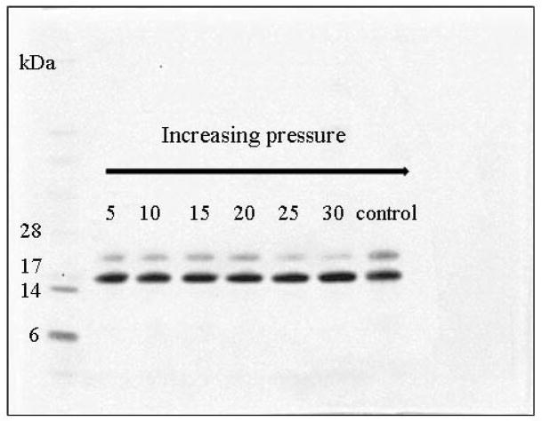
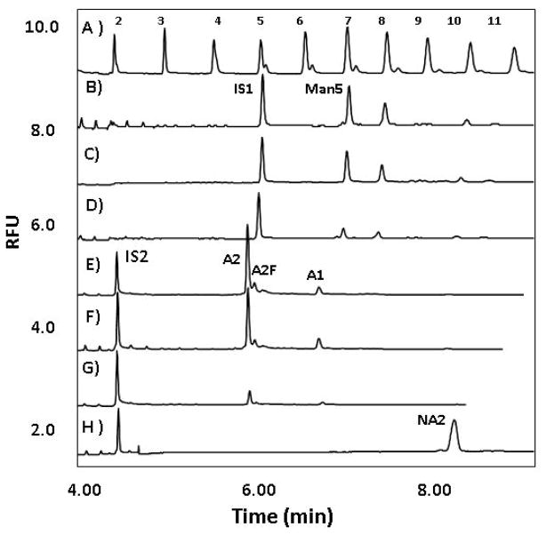
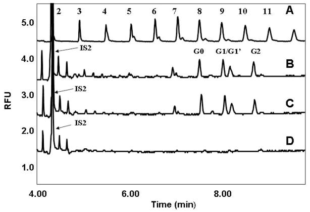
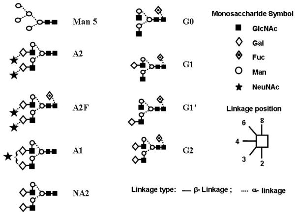
The standard, well-established sample preparation protocol to release N-linked glycans from glycoproteins for downstream analysis requires relatively long deglycosylation times (from several hours to overnight) and relatively high endoglycosidase concentration (from 1:250 to 1:500 enzyme:substrate molar ratio). In this paper, we significantly improve this standard protocol by the use of pressure-cycling technology (PCT) to increase the speed and decrease the relative amount of PNGase F during the release of N-linked glycans from denatured glycoproteins. With the application of pressure cycling from atmospheric to as high as 30 kpsi, >95% release of the asparagine-linked glycans from bovine ribonuclease B, human transferrin, and polyclonal human immunoglobulin was rapidly achieved in a few minutes using as low as 1:2500 enzyme:substrate molar ratio. The deglycosylation rate was first examined by SDS-PAGE at the protein level. The released glycans were then quantitated by capillary electrophoresis with laser induced fluorescence detection (CE-LIF). This new sample preparation protocol readily supports large-scale glycan analysis of biopharmaceuticals with rapid deglycosylation times.
Figures
Similar articles
-
Enzymatic removal of N-glycans by PNGase F coated magnetic microparticles.Electrophoresis. 2016 May;37(10):1264-9. doi: 10.1002/elps.201500575. Epub 2016 Mar 15. Electrophoresis. 2016. PMID: 26872155
-
A fast and convenient solid phase preparation method for releasing N-glycans from glycoproteins using trypsin- and peptide-N-glycosidase F (PNGase F)-impregnated polyacrylamide gels fabricated in a pipette tip.J Pharm Biomed Anal. 2020 Feb 5;179:112995. doi: 10.1016/j.jpba.2019.112995. Epub 2019 Nov 17. J Pharm Biomed Anal. 2020. PMID: 31767225
-
Release of bifidogenic N-glycans from native bovine colostrum proteins by an endo-β-N-acetylglucosaminidase.Enzyme Microb Technol. 2023 Jan;162:110138. doi: 10.1016/j.enzmictec.2022.110138. Epub 2022 Oct 4. Enzyme Microb Technol. 2023. PMID: 36252443
-
N-glycoproteomics in plants: perspectives and challenges.J Proteomics. 2011 Aug 12;74(8):1463-74. doi: 10.1016/j.jprot.2011.05.007. Epub 2011 May 12. J Proteomics. 2011. PMID: 21605711 Review.
-
The cytoplasmic peptide:N-glycanase (NGLY1) - Structure, expression and cellular functions.Gene. 2016 Feb 10;577(1):1-7. doi: 10.1016/j.gene.2015.11.021. Epub 2015 Nov 30. Gene. 2016. PMID: 26611529 Free PMC article. Review.
Cited by
-
Structural glycomic analyses at high sensitivity: a decade of progress.Annu Rev Anal Chem (Palo Alto Calif). 2013;6:237-65. doi: 10.1146/annurev-anchem-062012-092609. Epub 2013 Apr 3. Annu Rev Anal Chem (Palo Alto Calif). 2013. PMID: 23560930 Free PMC article. Review.
-
Comparative Proteomic Analysis of Cotton Fiber Development and Protein Extraction Method Comparison in Late Stage Fibers.Proteomes. 2016 Feb 3;4(1):7. doi: 10.3390/proteomes4010007. Proteomes. 2016. PMID: 28248216 Free PMC article.
-
N-deglycosylation targeting chimera (DGlyTAC): a strategy for immune checkpoint proteins inactivation by specifically removing N-glycan.Signal Transduct Target Ther. 2025 Apr 28;10(1):139. doi: 10.1038/s41392-025-02219-6. Signal Transduct Target Ther. 2025. PMID: 40289109 Free PMC article.
-
Spontaneous and site-specific immobilization of PNGase F via spy chemistry.RSC Adv. 2023 Sep 27;13(41):28493-28500. doi: 10.1039/d3ra04591a. eCollection 2023 Sep 26. RSC Adv. 2023. PMID: 37771922 Free PMC article.
-
Rapid and efficient protein digestion using trypsin-coated magnetic nanoparticles under pressure cycles.Proteomics. 2011 Jan;11(2):309-18. doi: 10.1002/pmic.201000378. Epub 2010 Dec 17. Proteomics. 2011. PMID: 21204257 Free PMC article.
References
-
- Varki A, Cummings R, Esko J, Freeze H, Stanley P, Bertozzi CR, Hart GW, Etzler ME. Essentials of Glycobiology. 2. Cold Spring Harbor Laboratory Press; Cold Spring Harbor, NY: 2009. - PubMed
-
- Jefferis R. Expert Opin Biol Ther. 2007;7:1401–1413. - PubMed
-
- Gennaro LA, Salas-Solano O. Anal Chem. 2008;80:3838–3845. - PubMed
-
- Li H, d’Anjou M. Curr Opin Biotechnol. 2009;20:678–684. - PubMed
-
- Townsend RR, Hotchkiss AT. Techniques in Glycobiology. Marcel Dekker; New York: 1997.
Publication types
MeSH terms
Substances
Grants and funding
LinkOut - more resources
Full Text Sources
Other Literature Sources
